We have a microfluidics research laboratory in the Institute of Medical Sciences on the Foresterhill campus.
Microfluidics is a state-of-the-art technology that offers tremendous experimental possibilities. Cells can be cultured in miniaturised culture chambers and their behaviour observed in real-time. For example, we are combining microfluidics and cell culture techniques to assess neurotoxicity in the neuromuscular system.
The laboratory designs and fabricates organ-on-chip devices for biomedical research. Our chips offer unprecedented experimental possibilities and solutions in areas ranging from neuroscience, myocardial infarction and cancer to thrombosis. We perform photolithography, soft lithography and mathematical modelling.
Each year we offer undergraduate projects, giving the students the opportunity of hands-on experience with this state of the art technology.
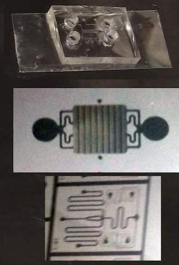
Some of our devices
Examples of cell images
_enhanced_reversed-1388x924.jpg)
Neurons (left) and muscle fibre (right) occupy different compartments on the same chip. Motor neurites pass through the thin cross channels (centre) to innervate muscle fibres.
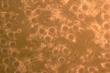
Beating cardiomyocytes (heart cells) grown for 3 days within our diffusion-based gradient generation microfluidic device. The regularly-spaced circular structures are part of the device.
Image credits: Dr Claudiu Giuraniuc; Amrita Debnath; Dr Claire Hetherington and Dr Abigail Dos Santos (two-chamber device); Saka et al., 2017 (diffusion-based device); Dr Victor Velecela, Kate Sword, and Art Dominey (cardiomyocytes).