PET imaging in the heart
PET Imaging in Drug Design & development (PET3D)
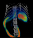
Figure: (a) PET/CT scan of a pre-clinical model's abdominal region. (b) Logo of the PET3D project.
Positron Emission Tomography (PET) imaging is most commonly used as a diagnostic technique - radioactive 'tracer' molecules are injected into patients and PET is used to observe the functioning of the body and identify signs of abnormalities, including heart conditions. PET3D is a €4million doctoral training scheme - coordinated by the University of Aberdeen - that will tackle a European shortage of scientists who can use PET Imaging for speeding up and reduce the cost of life-saving drug development.
Funded by the European Commission (H2020 Marie Skłodowska-Curie ITN-European Training Network)
Project team:
- Professor Matteo Zanda (Research scientist, University of Aberdeen)
- Dr Sergio Dall'Angelo (Research scientist, University of Aberdeen)