Scientists at the University of Aberdeen and NHS Grampian have been awarded £350,000 of Scottish Government funding to investigate a new way to scan brain tumours.
Funded by a Chief Scientist Office Translational Clinical Studies grant, the team will use Aberdeen-designed, Field Cycling Imaging (FCI) to generate never-before seen images of glioblastoma brain tumours.
Glioblastoma is the most common and aggressive type of brain tumour with over 3,000 new patients in the UK diagnosed each year. Half of all patients die within 15 months of diagnosis even after extensive surgery, radiotherapy and chemotherapy.
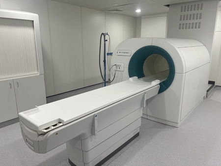
Field cycling imaging (FCI) is a new and specialist type of low-field MRI scan pioneered in Aberdeen. The FCI scanner follows in the footsteps of the full body MRI scanner, also invented at the University around 50 years ago which has gone on to save millions of lives around the world. The FCI derives from MRI but can work at low and ultra-low magnetic fields which means it is capable of seeing how organs are affected by diseases in ways that were previously not possible.
While similar to MRI, in that MRI uses strong magnetic fields and radio waves to produce detailed images of the inside of the body, the FCI scanner can vary the strength of the magnetic field during the patient’s scan. This means the FCI acts like multiple scanners in one and can extract more information about the tissues.
A further benefit of this new technology is that it can detect tumours without having to inject dye into the body, known as contrast agents, which have been associated with kidney damage and allergic reactions in some patients.
The Aberdeen scanner is the only one of its type used in patients anywhere in the world.
The team of doctors and scientists involved in this project will scan glioblastoma patients undergoing chemotherapy after surgery and chemoradiotherapy.
They hope to show that, unlike conventional MRI scans, FCI can tell the difference between tumour growth, known as progression, and ‘pseudo-progression’ which looks like tumour but is not cancerous tissue.
If they can distinguish pseudo-progression from true progression this could improve care and quality of life in future patients.
Professor Anne Kiltie, Friends of ANCHOR Chair in Clinical Oncology at the University of Aberdeen, who is leading the study said: “We already have evidence that FCI is effective in detecting tumours in breast tissue and brain damage in patients following a stroke.
“Applying this exciting new technology to glioblastoma patients could give us a much more accurate and detailed picture of what is going on in their brain. If we can detect true tumour progression early, we can swap the patient to a potentially more beneficial type of chemotherapy. Also, being able to verify that a patient has pseudo-progression will prevent effective chemotherapy being stopped too early, because it was thought that the tumour has progressed, thus worsening prognosis.
“Providing certainty will also reduce anxiety for both patients and relatives and improve the quality of life of patients.
“Importantly, having a reliable method to identify progressive disease will allow development and more precise evaluation of emerging potential treatments. This is of particular importance as patients currently have a limited choice of treatments for combatting their cancer.
“Ultimately this study and related future work will improve quality, effectiveness and healthcare cost-effectiveness in the treatment of glioblastoma patients across Scotland and beyond.”
Sarah-Jane Hogg, chief executive at Friends of ANCHOR, added: “This is a really promising development and another example of the pioneering work coming out of the University of Aberdeen.
“Professor Kiltie’s role at the University is fully funded by Friends of ANCHOR through our Dream Big appeal, and our thanks go to our donors and fundraisers for the part they’ve played in supporting this work.”