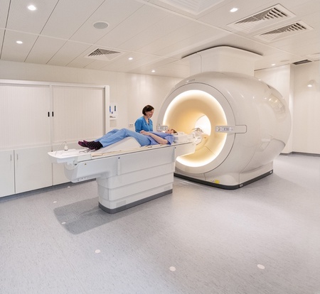
Anyone who has experienced an MRI scan will be familiar with the strange clunking and whirring of the large tube-shaped machines which are a fixture of hospitals around the world.
A strong magnet and radio waves are used to gather signals from the body, and a computer turns those signals into images that doctors can study.
Today we take for granted this technology to enable doctors to see inside the human body. But the story of the first full-body MRI scanner began in a small laboratory at the University of Aberdeen.
In the early 1980s Aberdeen researchers – working on a shoestring budget – built and tested a machine which could detect cancer and other changes in body tissue. It was a breakthrough which shaped modern medical treatment. Today the University continues to lead advances in medical imaging through both Field‑Cycling Imaging and Low-Field MRI. Read our collection of three long-read articles to find out how the MRI story began and how the University could lead the next chapter.
The first full-body MRI scanner
In the early 1960s, a young researcher named John Mallard was developing his research interest in the magnetic properties of electrons in tissue samples. He made the exciting observation that there were differences between normal tissues and tumours.
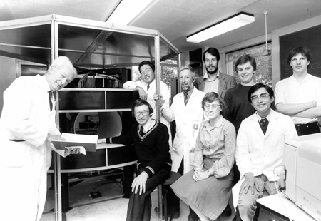
In 1965 he moved from London to Aberdeen where he led what he would later describe as "a small team of half-mad scientists" who were instrumental in the development of MRI technology which would change the course of medical history forever.
Read about the Aberdeen MRI pioneers.
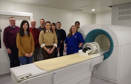
How Aberdeen is shaping the future of medical imaging
Four decades on, University scientists are building on that legacy exploring new forms of imaging that could help clinicians detect disease earlier, understand it better and tailor treatments more precisely.
While modern MRI scanners produce incredibly detailed images, Aberdeen researchers are now asking a different question: what information might be hidden beyond the image itself?
That question is at the heart of a new approach known as Field-Cycling Imaging (FCI), a technology being developed and explored at the University of Aberdeen.
Read more about how FCI could contribute to the future of medical imaging.
How Low-Field MRI is building on Aberdeen’s pioneering legacy
A new research team is opening another chapter in Aberdeen's MRI story. Instead of pushing MRI to ever‑higher magnetic strengths, they’re exploring what becomes possible when those strengths are deliberately lowered.
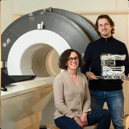
Their work on Low‑Field MRI could transform cost, access and patient experience at a time when half the world still lacks advanced diagnostic imaging. Read the full story here.