Continuing a tradition of discovery:
How Low-Field MRI is building on Aberdeen’s pioneering legacy

For more than four decades the University of Aberdeen has played a distinctive role in the global development of magnetic resonance imaging (MRI).
From the pioneering 1980s research team that built the world’s first full-body MRI scanner on a shoestring budget to today’s internationally recognised work in Field-Cycling Imaging (FCI), the city has helped shape how MRI is understood and applied in medicine.
Now, that legacy is entering a new chapter. A newly established research team is once again pushing the boundaries of MRI – not by increasing magnetic field strength, but by exploring what becomes possible when it is deliberately reduced.
Working alongside Aberdeen’s existing research strengths, the team is investigating how Low-Field MRI could transform the cost, accessibility and patient experience of medical imaging – a crucial step when around half the world’s population still cannot access advanced diagnostic imaging.
A city shaped by MRI innovation

Aberdeen’s relationship with MRI stretches back to the early 1980s, when researchers were still working out how magnetic resonance could be transformed into a practical medical imaging tool. At that time, powerful superconducting magnets were not yet available, and early systems operated at what would now be considered very low magnetic field strengths.
“People often forget that MRI started at low field. That wasn’t a choice – it was simply all that existed,” says Dr Mathieu Sarracanie, a Magnetic Resonance Physicist and one half of a research partnership brought to the University to reimagine MRI from a new perspective.
Over time, advances in magnet technology pushed MRI towards progressively higher field strengths. These systems delivered remarkable image quality but also became larger, more expensive and increasingly complex to build and operate.
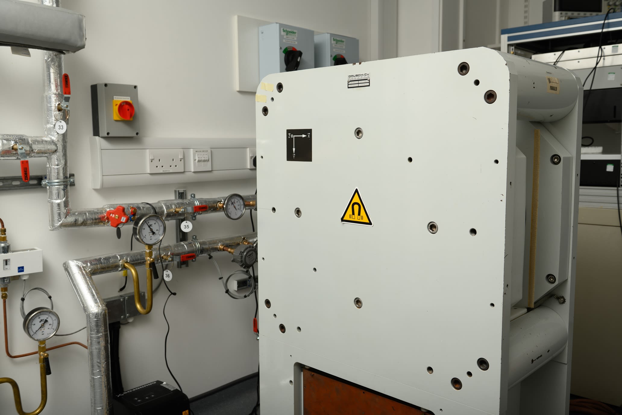
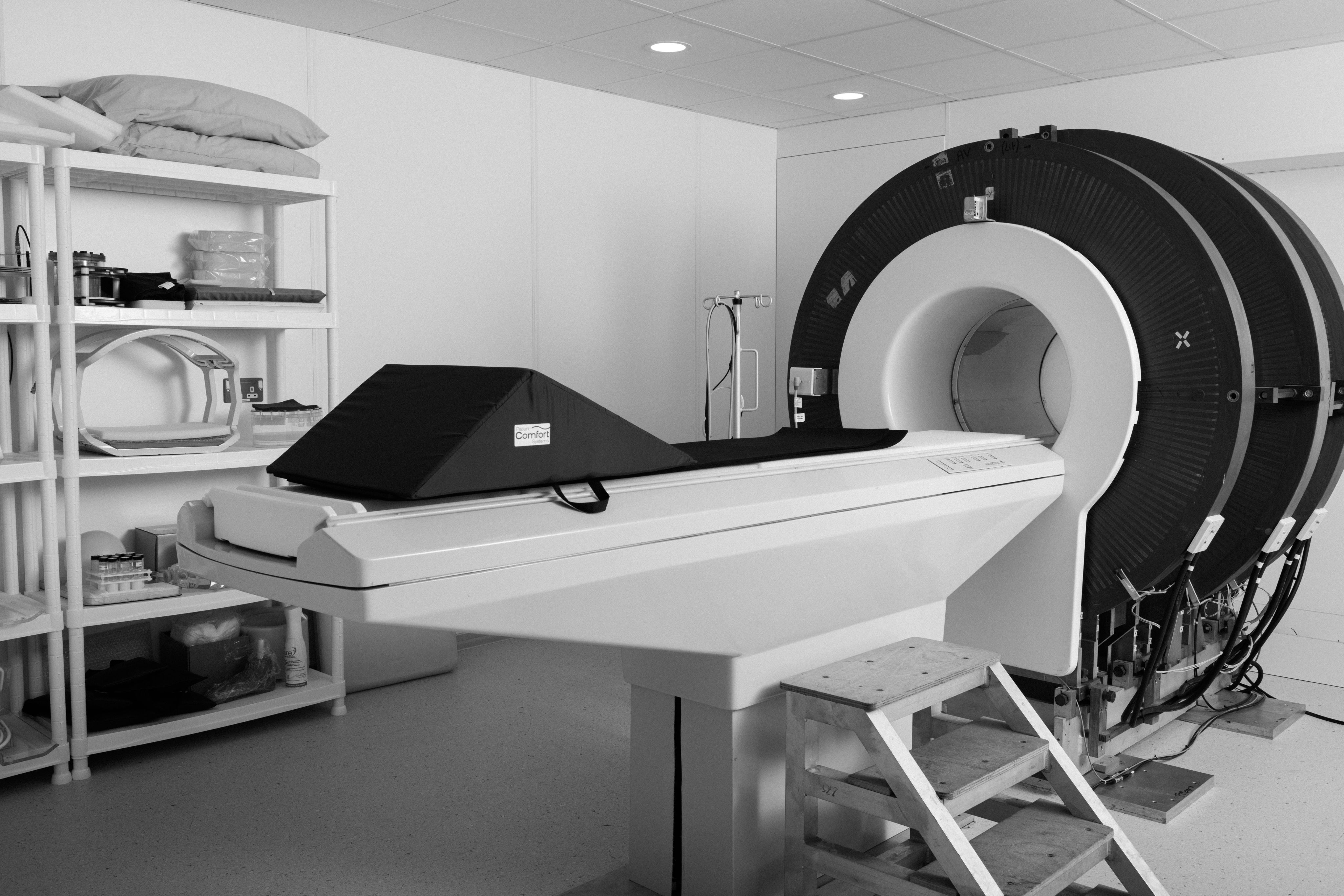
As MRI technology matured, Aberdeen continued to lead innovation, most notably through the development of Field-Cycling Imaging (FCI) . This unique technique allows the magnetic field to be actively varied during a single scan, revealing new forms of tissue contrast that cannot be captured using conventional MRI.
The research is providing new insights into how tissues behave across different magnetic fields, with the potential to improve early disease detection, distinguish between similar conditions, and monitor how illnesses respond to treatment. However, like traditional MRI scanners, field-cycling systems are not designed to be small, low-cost or portable.
Dr Sarracanie, together with research partner Dr Najat Salameh, aims to develop clinically useful MRI technologies that address these challenges by returning to where Aberdeen’s MRI journey first began – innovation built around smaller, less powerful magnets.

Bringing a new approach to Aberdeen

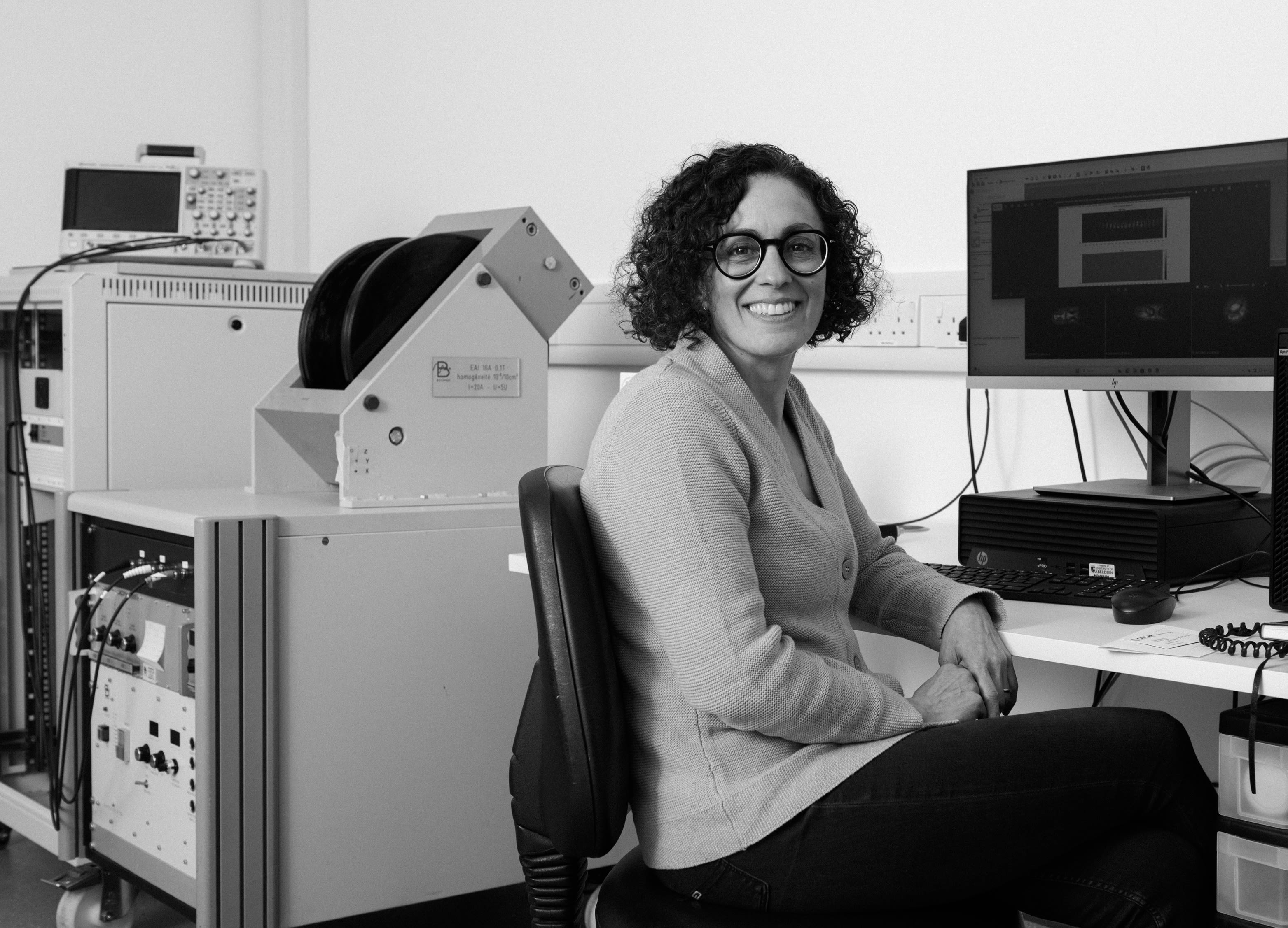
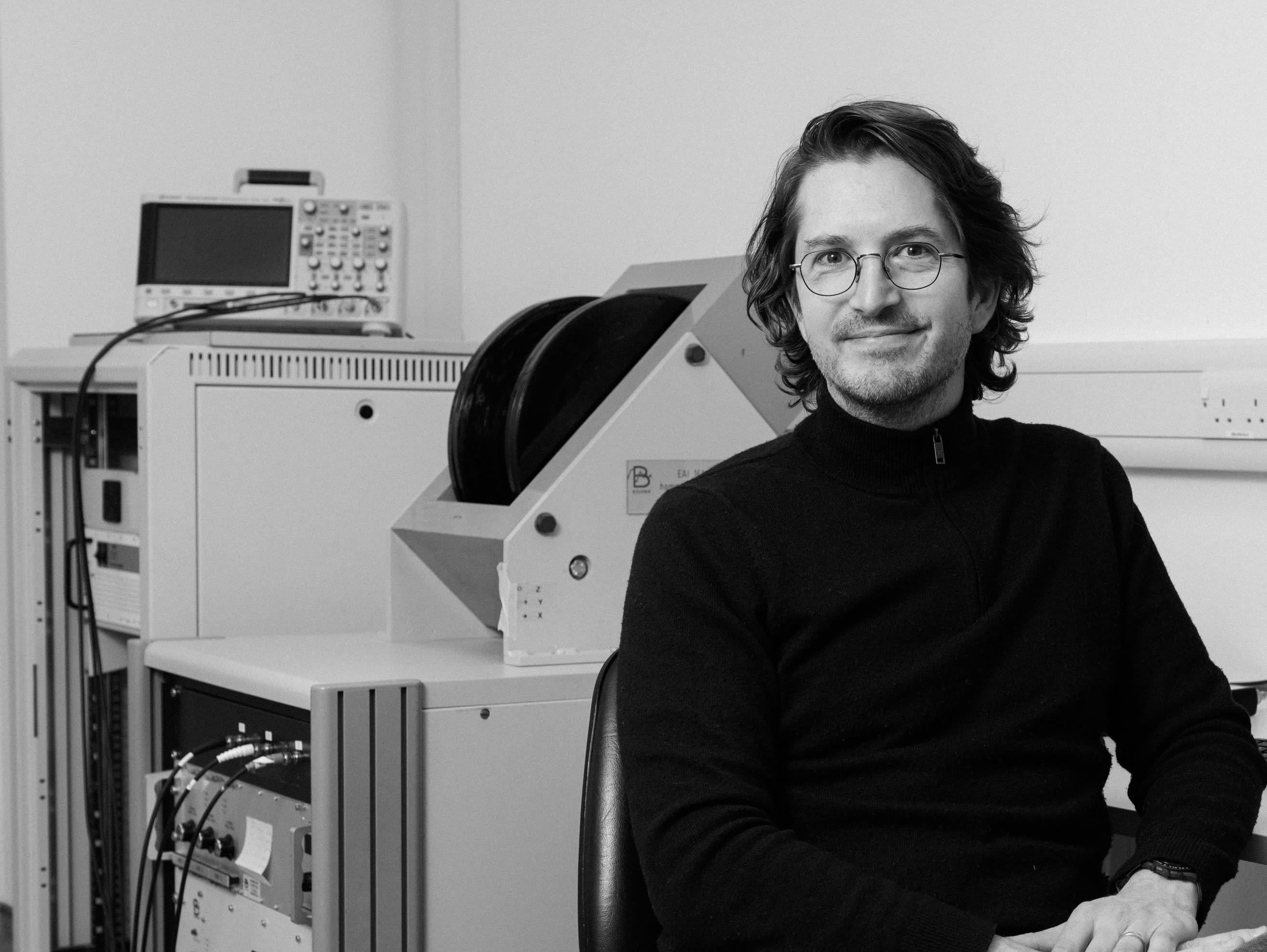
When Dr Sarracanie and Dr Najat Salameh arrived in Aberdeen in 2023, they brought with them years of international experience from research posts in France, Switzerland, and the United States, along with custom-built prototype scanners developed through major competitive grants. These were painstakingly dismantled and reassembled in their new home in the Biomedical Physics Building.
Their focus is fundamentally different from field-cycling MRI.
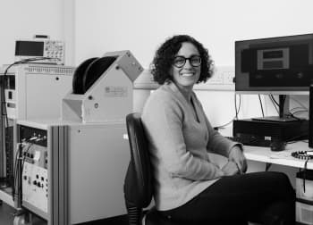
“We work with one magnetic field,” explains Dr Salameh. “It’s a low magnetic field, and it stays there during the scan. We don’t cycle the field.”
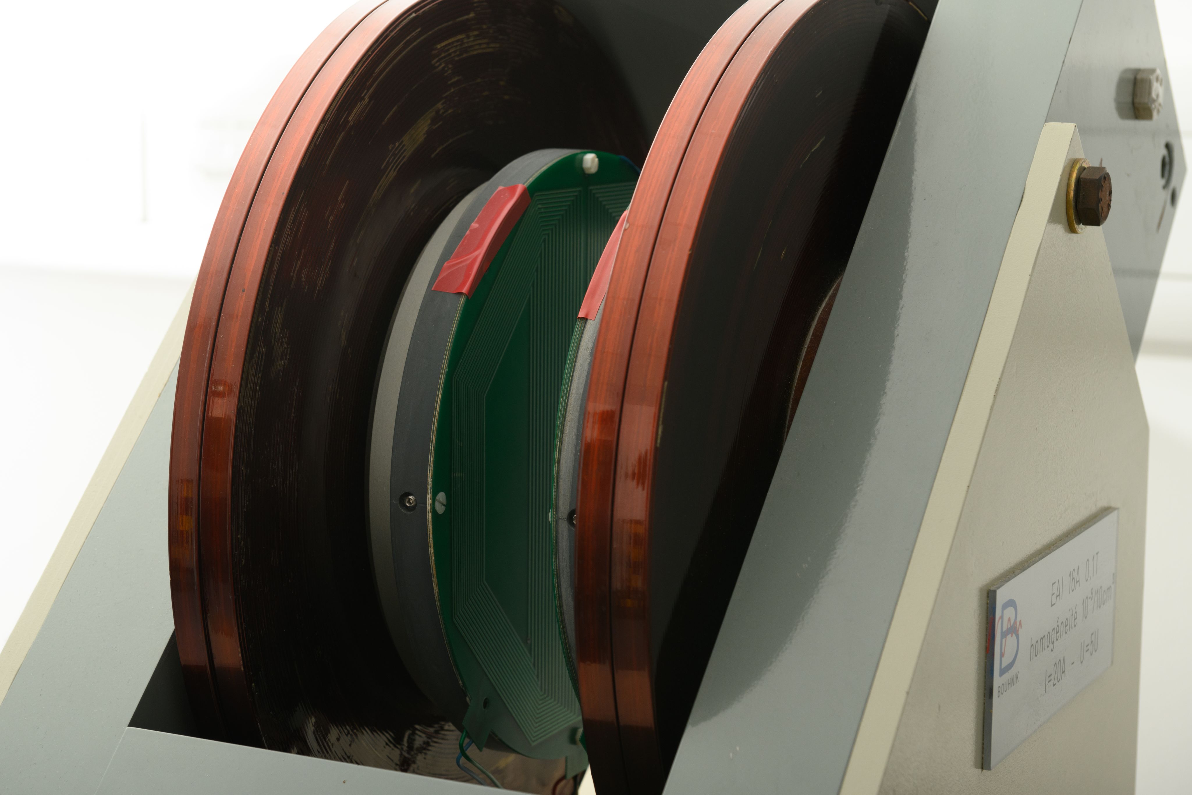
This seemingly simple distinction has profound implications. By keeping the magnetic field fixed, the team can focus on optimising image quality, speed, and robustness – while stripping away much of the expensive hardware that dominates conventional MRI systems.
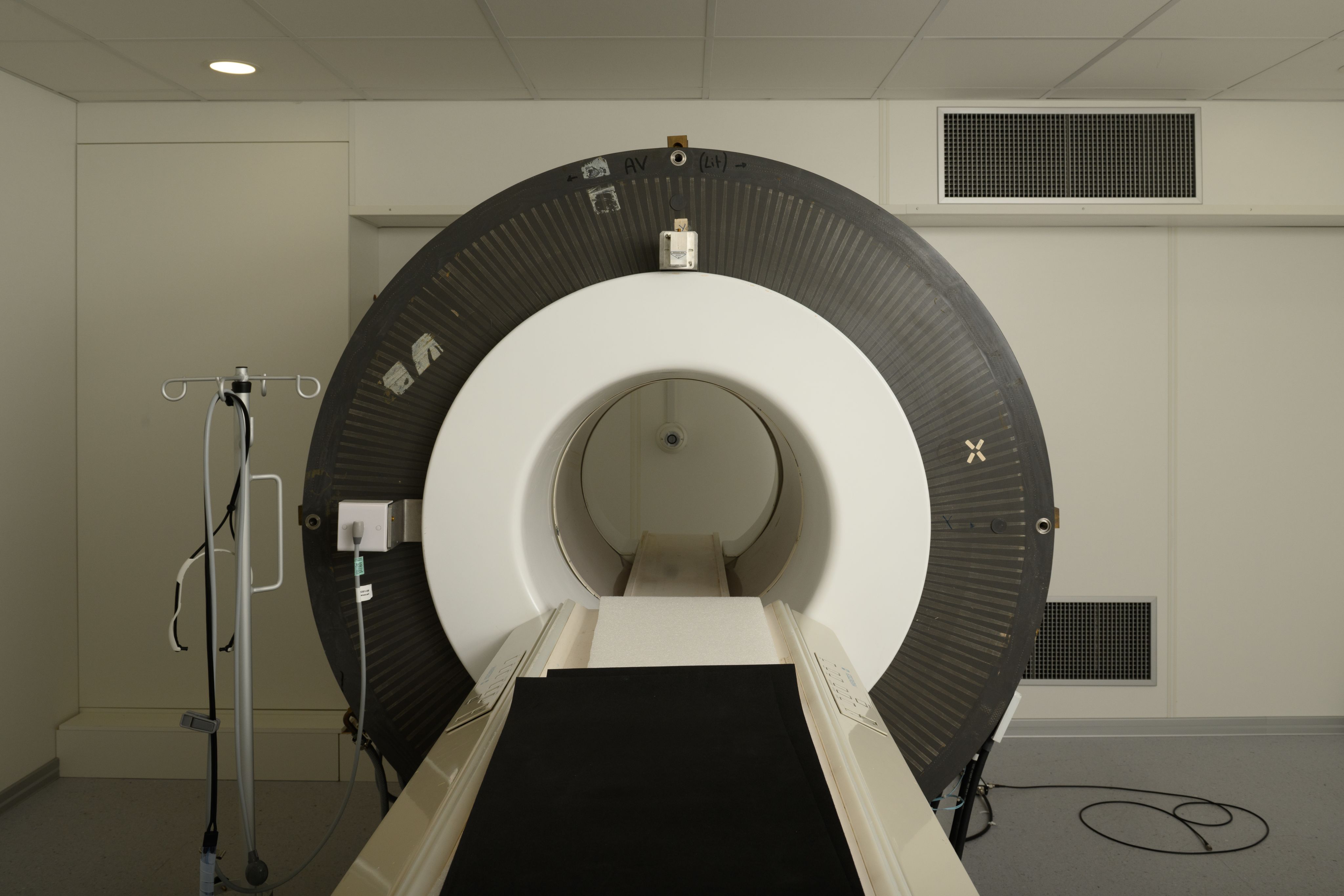
The result is low-field, fixed-field MRI scanners that are smaller, potentially portable, and far less costly to build and operate.

Dr Mathieu Sarracanie
Dr Mathieu Sarracanie
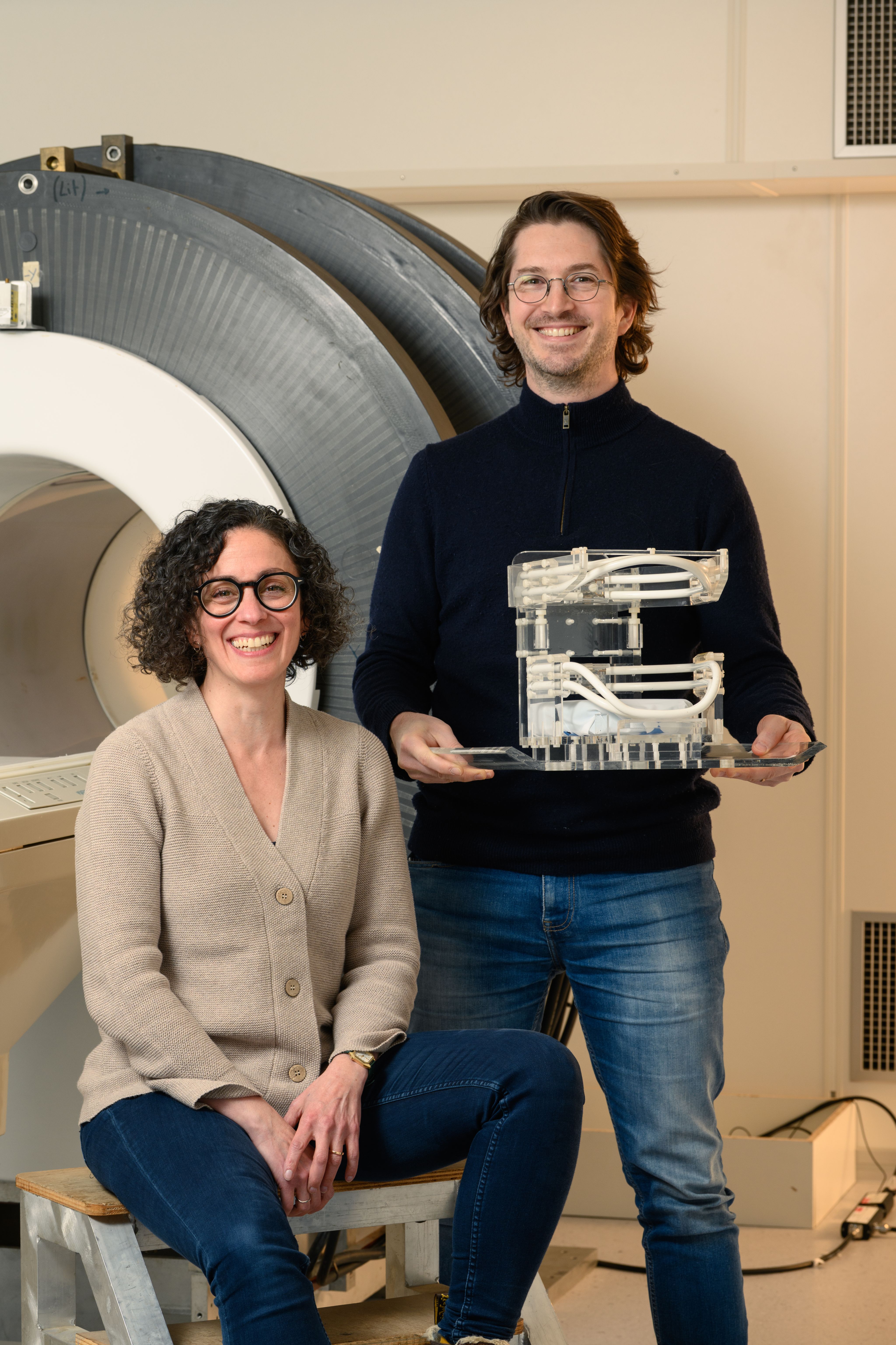
Dr Najat Salameh (left) and Dr Mathieu Sarracanie
Dr Najat Salameh (left) and Dr Mathieu Sarracanie
Lower magnetic field – bigger challenges

At its core, MRI relies on strong magnetic fields to generate signals from the body. The stronger the field, the stronger the signal, which is why modern hospital scanners typically operate at 1.5 or 3 Tesla.
Low-field MRI deliberately operates far below these levels bringing a new set of challenges.
“At low magnetic field, your life is harder,” says Dr Sarracanie. “You have less signal, more noise, and everything becomes more complicated.
“It means that we have to be extra creative in maximizing the signal we can collect and reducing that noise so that the image quality is good for clinical diagnostics.”
It’s a process that would have been unthinkable even 10 years ago because of the difficulties in obtaining clinically-relevant images so why pursue it at all?
Dr Sarracanie points to a favourite quote from John F Kennedy’s famous ‘We choose to go to the moon’ speech: we do things ‘not because they are easy, but because they are hard’.
“More than half the world’s population still does not have access to MRI at all and 80% struggle to have access to MRI. That’s something we want to change but to do so a different approach is needed.
“What really locks MRI into being big, expensive, and inaccessible is the magnet. To introduce options which are scalable, portable, and widely available, you have to reduce the magnetic field.”
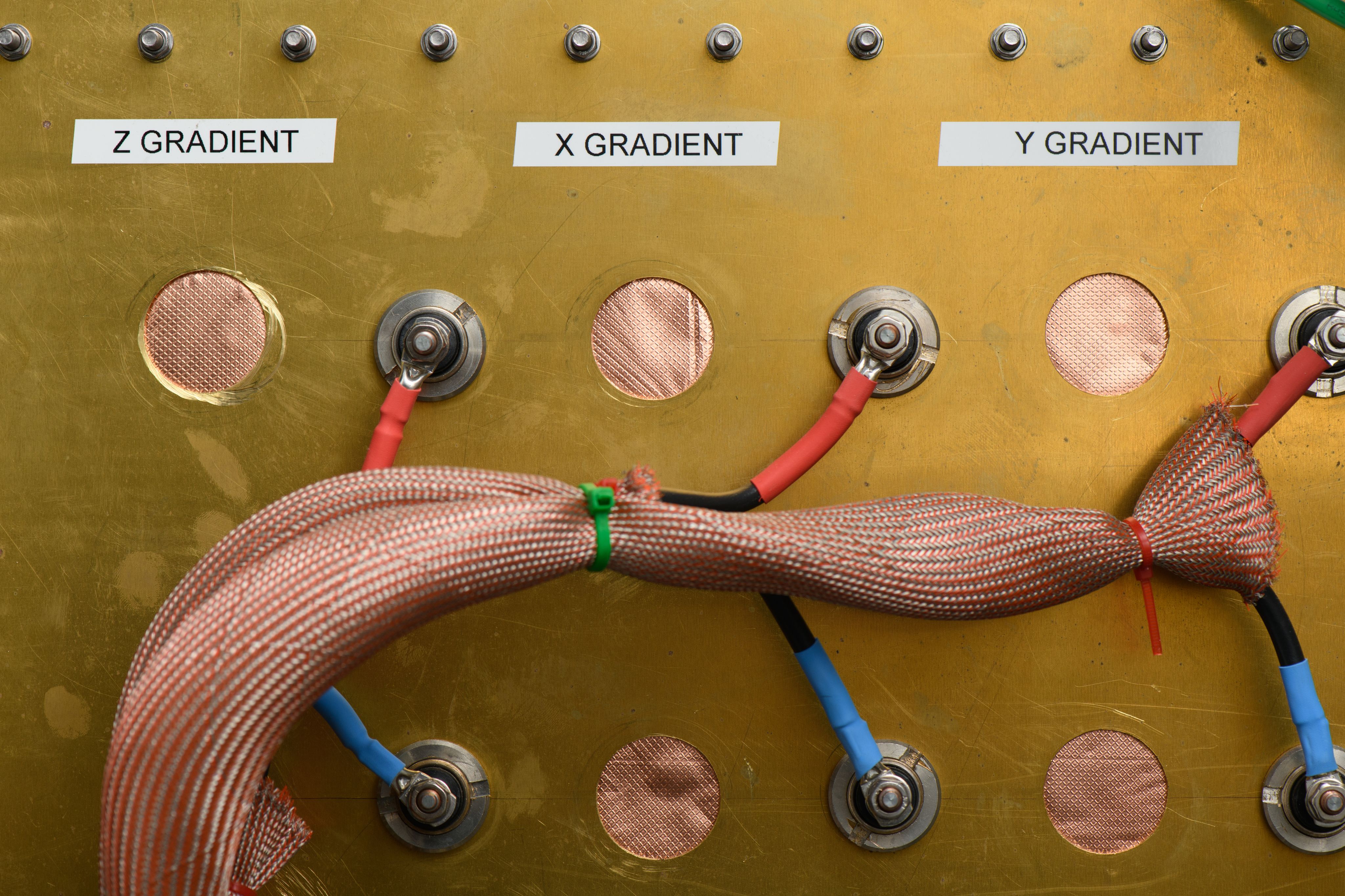
Lowering field strengths allow for simpler magnet designs, reduced shielding, lower power requirements, and greater flexibility in the size and shape of scanners.
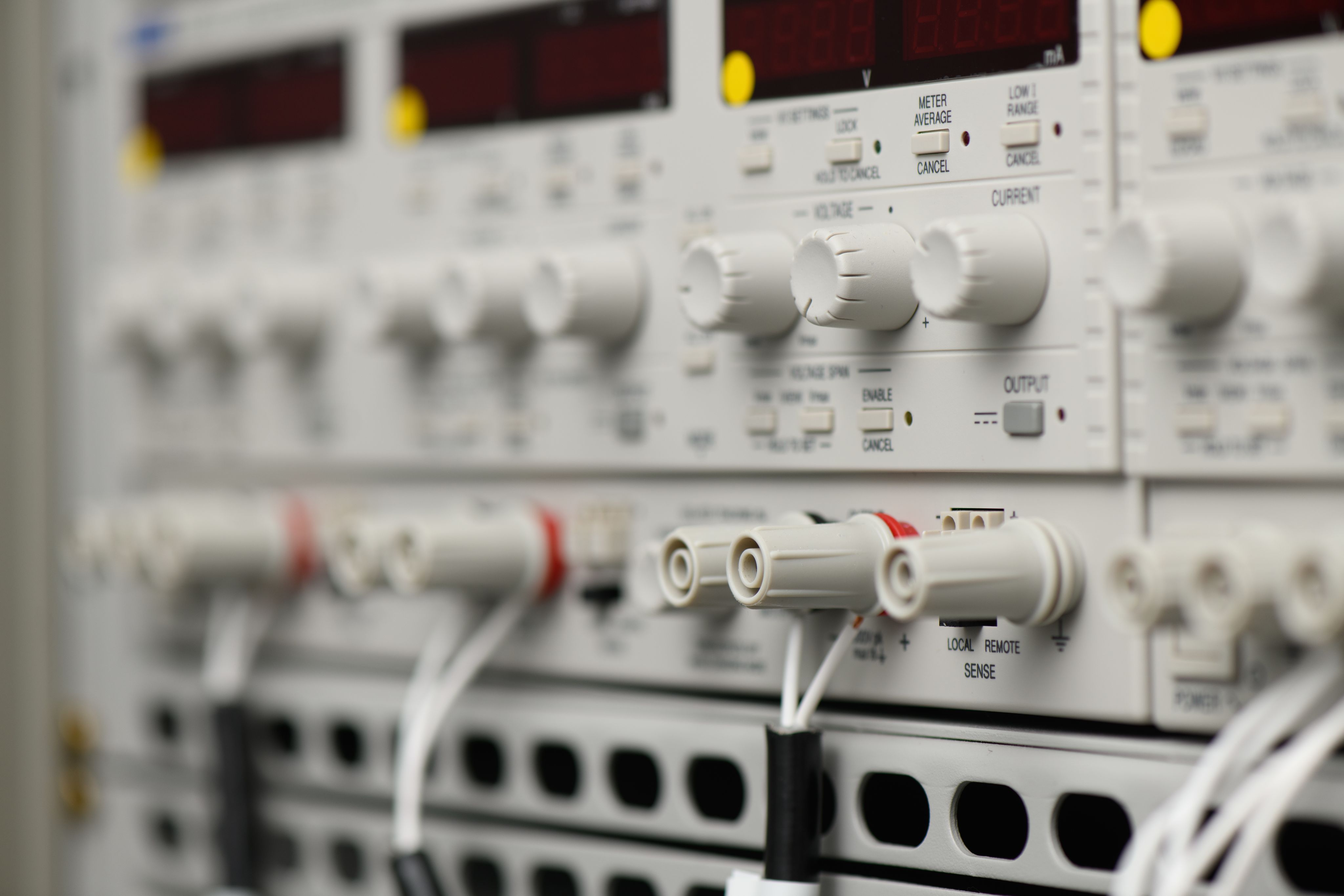
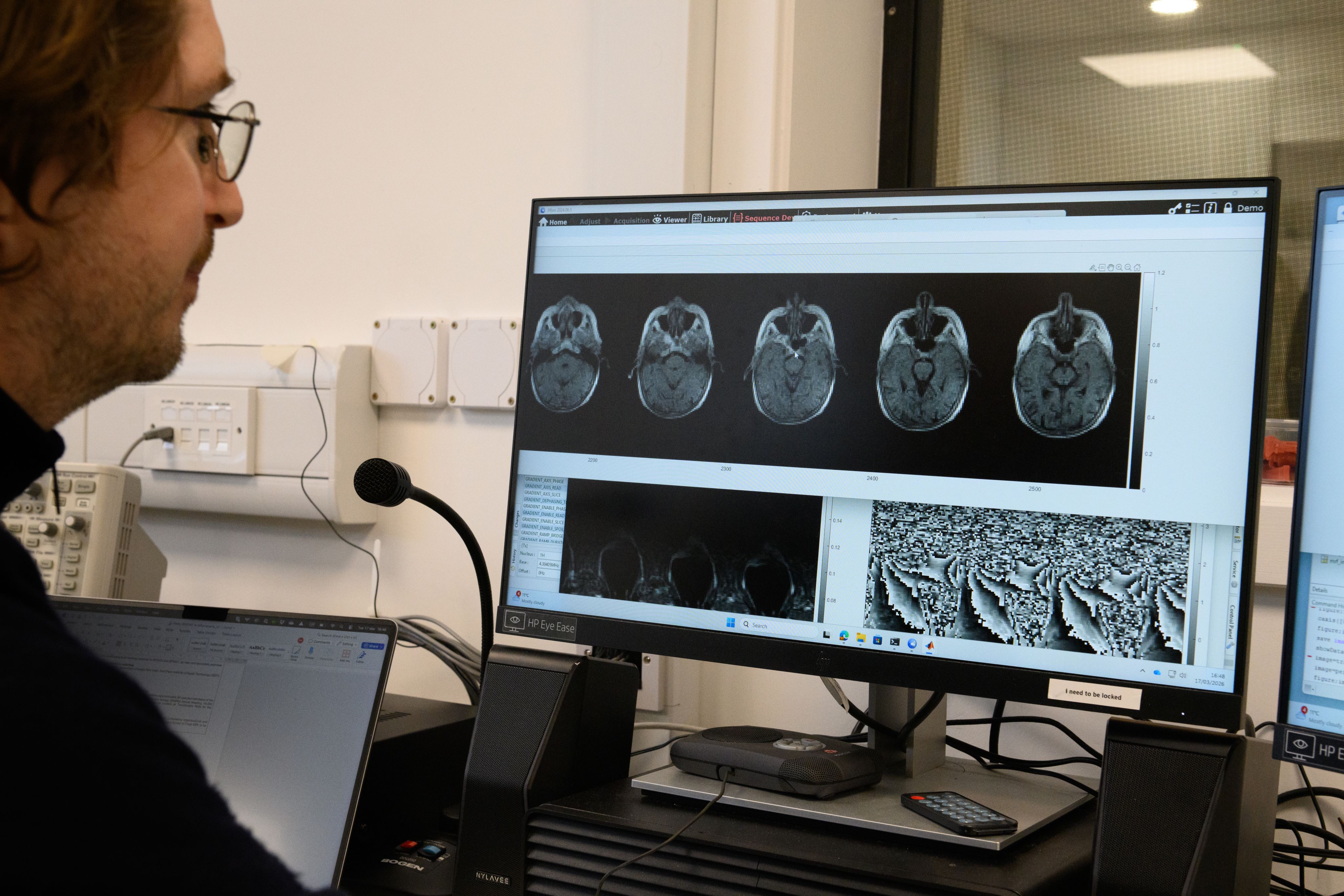
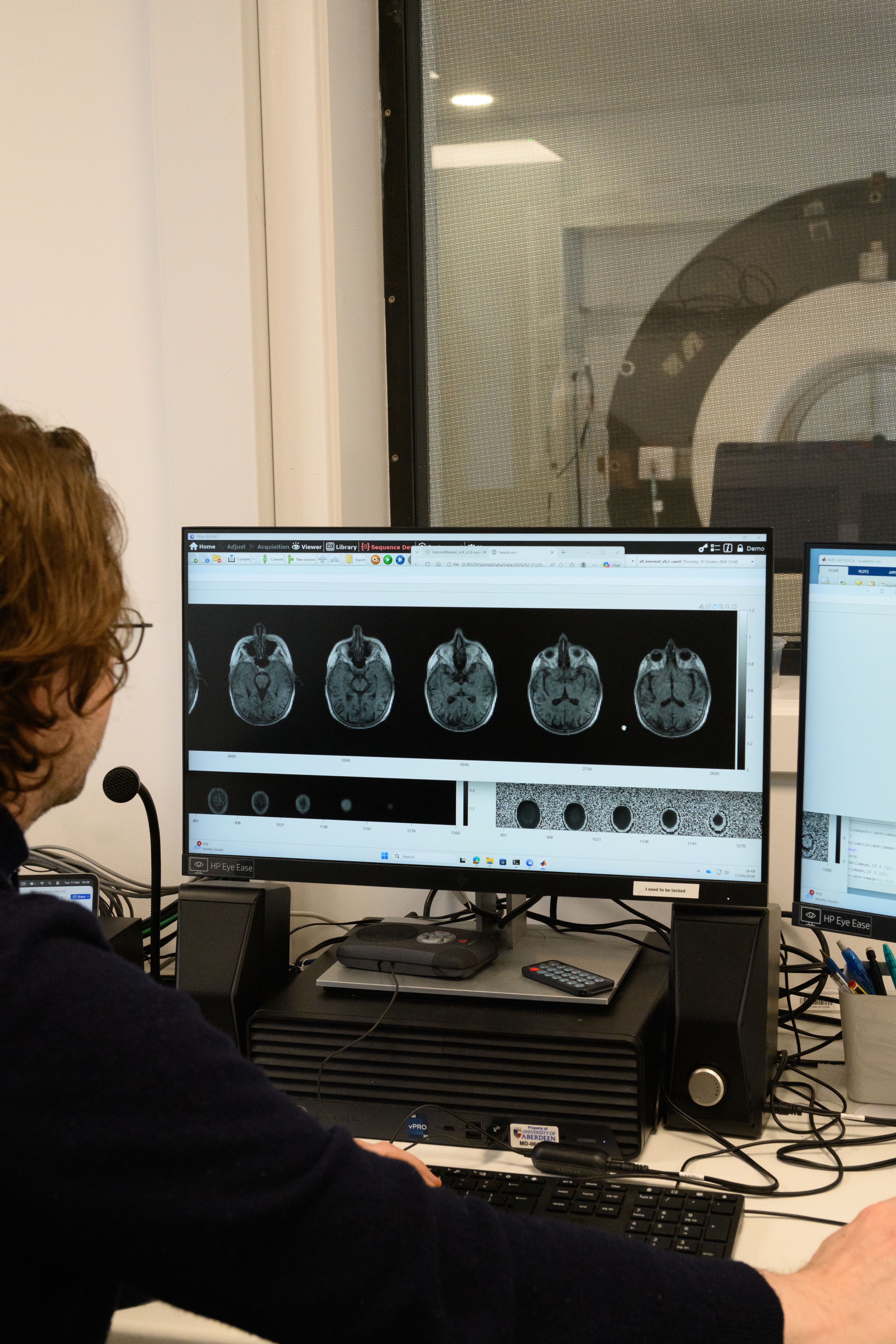
Using modern electronics, reconstruction algorithms, and signal processing, the team has shown that clinically useful images can still be achieved.
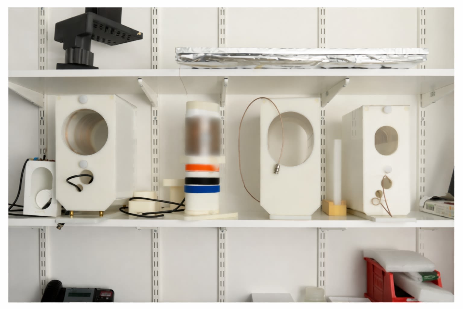
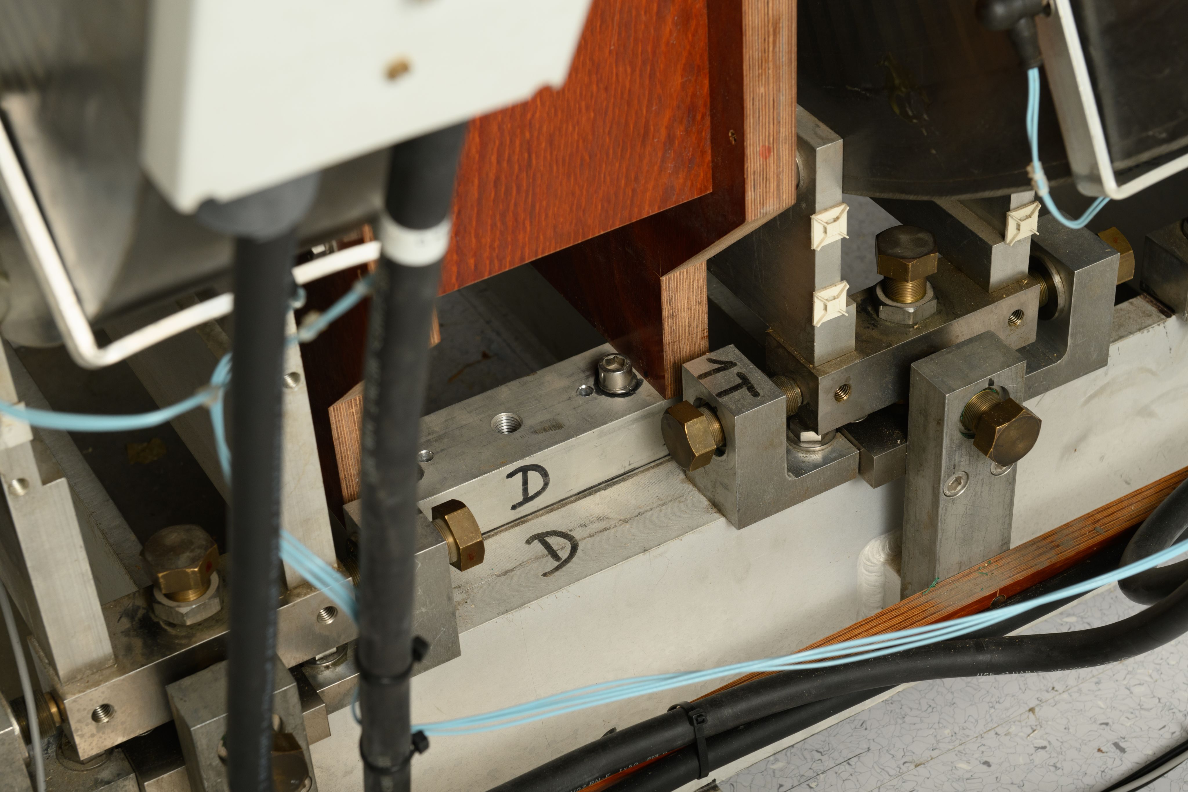
They’ve now assembled a range of prototype scanners which will soon be used on their first patients.
These include a full-body scanner as well as smaller systems designed specifically for extremities such as hands, the head and the abdomen.
Benefits for clinicians and patients

The potential benefits of Low-Field MRI extend well beyond a simplified build process.
For clinicians, low-field MRI could offer new tools for triage, screening, and follow-up, reducing pressure on overstretched high-field scanners. Faster access to imaging means faster decisions — even if a high-field scan is still needed later.
For patients, the experience itself may change.
Lower magnetic fields allow for quieter scanners, wider openings, and designs that avoid the long, narrow tunnels many people find claustrophobic. Dedicated scanners for hands, joints, or heads could allow patients to sit comfortably, communicate with staff, and even have a family member present during the scan.
Even in well-resourced healthcare systems, patients can wait months for scans. Low-field MRI offers a way to rethink how and where imaging happens – in hospitals, community settings, and potentially even at the bedside.
Low-field MRI also improves compatibility with implants, prosthetics, and other metallic objects, potentially widening access for patients who are currently excluded from conventional MRI.
“From those with cardiac pacemakers, implantable cardioverter-defibrillators (ICDs) and cochlear implants to patients with prostheses, MRI accessibility is far from universal even where good provision exists,” says Dr Salameh.
“As magnetic field strength decreases, interactions with the surrounding magnetic environment are reduced. This opens opportunities for imaging patients who are currently excluded from conventional MRI and for future interventional applications.”

Complementing field-cycling MRI, not competing with it

Crucially, low-field MRI in Aberdeen is not a rival to Field-Cycling Imaging (FCI), the two approaches are designed to work together.
“Field-cycling is about discovering new contrast – it’s a new imaging modality in its own right,” says Dr Sarracanie.
He compares FCI to viewing disease through a rainbow created from white light. Just as a prism separates white light into its component colours, FCI allows researchers to observe how tissues behave across a wide range of magnetic fields, revealing biological information that has previously been hidden.
Low-field MRI, by contrast, focuses on a single, carefully selected point within that spectrum. Rather than capturing every possible signal, it concentrates on one magnetic field that provides the most clinically useful information, delivering that insight consistently across the whole scan.
“Some diseases may benefit from seeing the full spectrum, while others may only require one specific signal,” says Dr Sarracanie. “The goal is to use the right part of the spectrum for the right clinical question.”

Looking ahead: carrying Aberdeen’s MRI legacy forward

Aberdeen’s MRI story has been defined by a willingness to challenge convention, and the development of Low-Field MRI represents the next step in that tradition.
“There is a real sense that we are continuing something that began here,” says Dr Salameh. “The early pioneers assembled the original Mark 1 scanner in a University laboratory, turning ambitious ideas into a machine capable of scanning patients and producing clinically useful images.
“But they did not stop at proving the concept. They continually refined their work – improving image quality and enhancing the patient experience as they learned from each scan.
“Four decades on, we are following that same philosophy, embracing their spirit of invention and applying it to the needs of modern healthcare.”