The "small team of half-mad scientists" who changed the course of medical history
The University pays tribute to Professor John Mallard, who died in February 2021, and his team of MRI pioneers

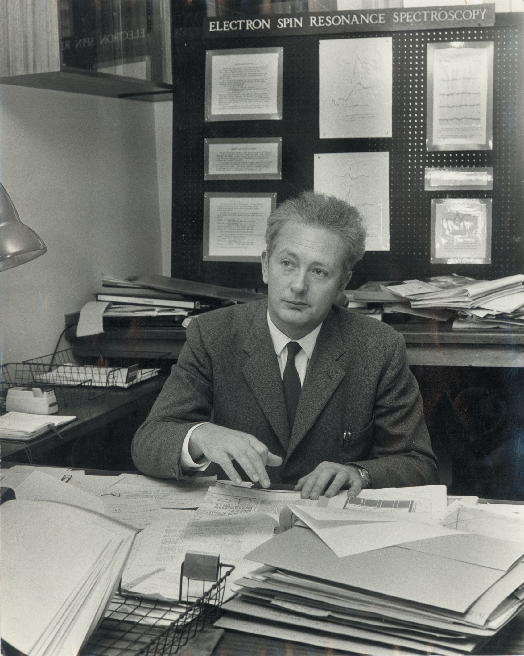
In the early 1960s, a young researcher named John Mallard was developing his research interest in the magnetic properties of electrons in tissue samples. He made the exciting observation that there were differences between normal tissues and tumours.
In 1965 he moved from London to Aberdeen where he led what he would later describe as "a small team of half-mad scientists" who were instrumental in the development of MRI technology which would change the course of medical history forever.
Following his death aged 94, the University looks back on his remarkable career and the extraordinary achievements of his team.
Seeing the big picture
Mallard became the University’s first Chair of Medical physics and appointed Jim Hutchison to work on a project exploring the possible medical uses of magnetic resonance.
After a number of false starts in their research, a breakthrough American study showed that magnetic resonance could distinguish between normal and cancerous tissue. American chemist Paul Lauterbur then proposed a technique to produce images using magnetic resonance.
Mallard and Hutchison decided to bring these elements together and in 1974 they obtained the first ever MRI picture of a mouse using a desktop scanner which they had constructed, although it took several hours to produce the image.
Mallard immediately saw the ‘big picture’ and where other research teams around the world sought to increase the technology on a smaller scale, obtaining images of fingers for example, he felt its true medical use lay in the ability to obtain whole body scans. The technology he would drive needed to be able to scan the organs of the body from the brain to the liver and kidneys and for that to happen, a much larger scaling up was needed.
But convincing funders to back his ideas proved a battle and after many trips between Aberdeen and London, he eventually persuaded the Medical Research Council to support the team’s endeavours.
The researchers, who by this time had recruited chief technician Eddie Stevenson from the Medical Physics workshop, then set about design and construction.

Development on a shoestring budget
The Mark-1 scanner they created was put together on a shoestring budget of just £30,000, which also had to stretch to cover the staff time.
Hutchison had designed the magnet and it was delivered to Aberdeen from Oxford in 1977. This then required large radio-frequency coils around it so the team set about building these from central heating pipes and utilised other materials available to them at low-cost, including a tube from a children’s play park.
The magnet had also arrived without any form of cooling system and after running it for an hour, became so hot that it could fry an egg.
University PhD students were enlisted to develop a water cooling system and although successful, in the early days this was plagued with leaks and the team spent a considerable amount of time dealing with dripping water.


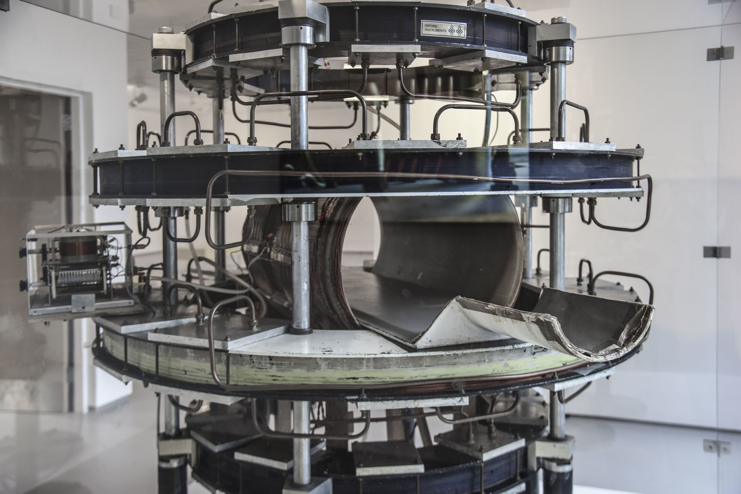
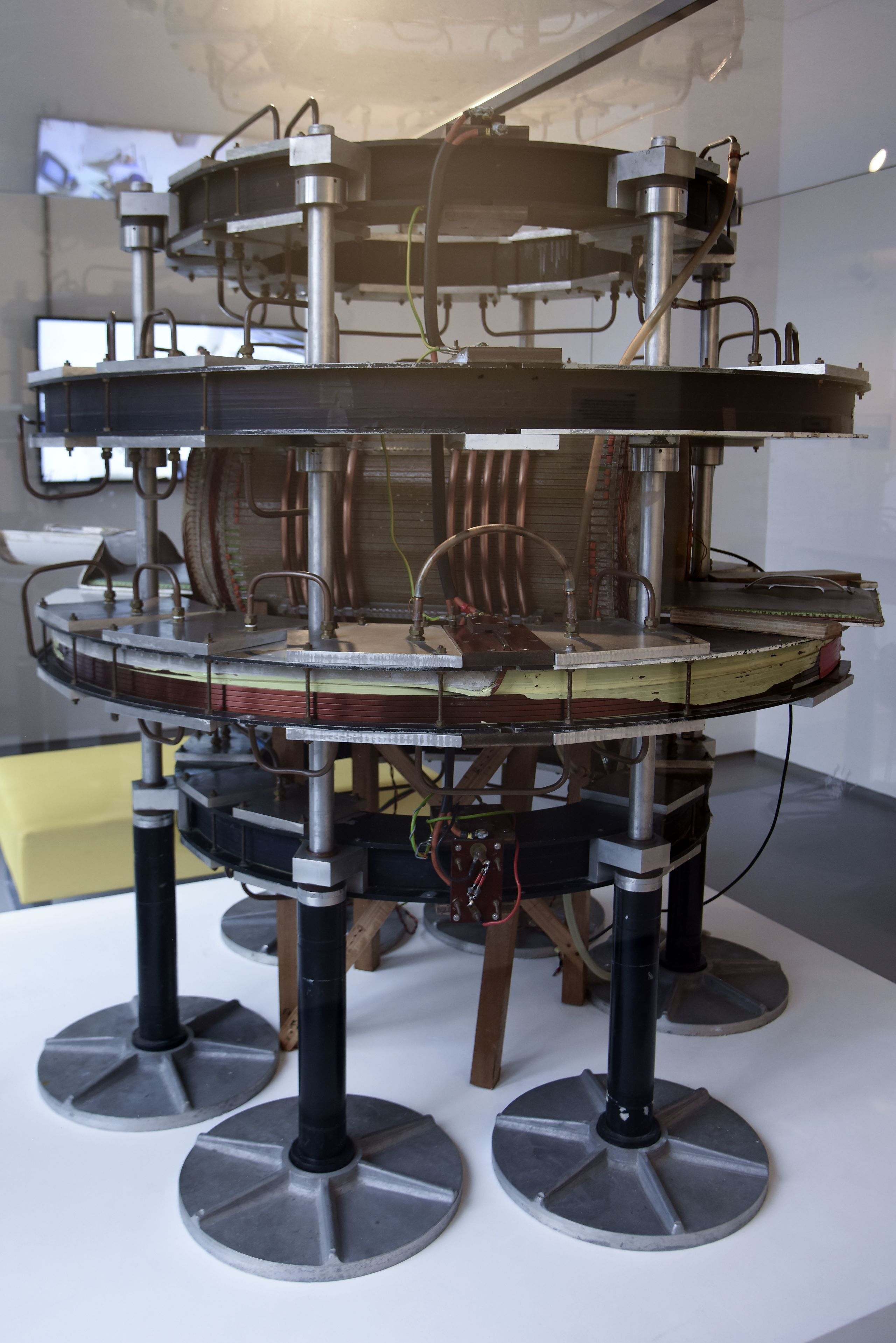
The first magnet before the MK-1 equipment
The first magnet before the MK-1 equipment

The ‘spin warp’ breakthrough
Despite progress, the team were still unable to reconstruct images which were diagnostically useful with blobby and grainy pictures returned.
They had to overcome the natural movement within the body, such as the beating of the heart, and develop a system which could ‘freeze’ the images.
Their ‘eureka moment’ came following the appointment of a young American postdoctoral scientist named Bill Edelstein.
Using their combined expertise the team came up with the solution that was to prove the breakthrough that MRI needed – the spin-warp imaging pulse sequence.
It was hoped the technique could eliminate the stripes and squiggles, known as artefacts, caused by involuntary movement of the body.
To test the theory, the team worked through the night and in April 1980, Hutchison took his place as ‘patient’ within the scanner while Edelstein programmed and operated the machine from a computer room down the corridor with cables stretched between them.
In that instant, medical history was made. Spin warp provided high quality images of the body that were by far the best in the world at that time and is still widely used today.

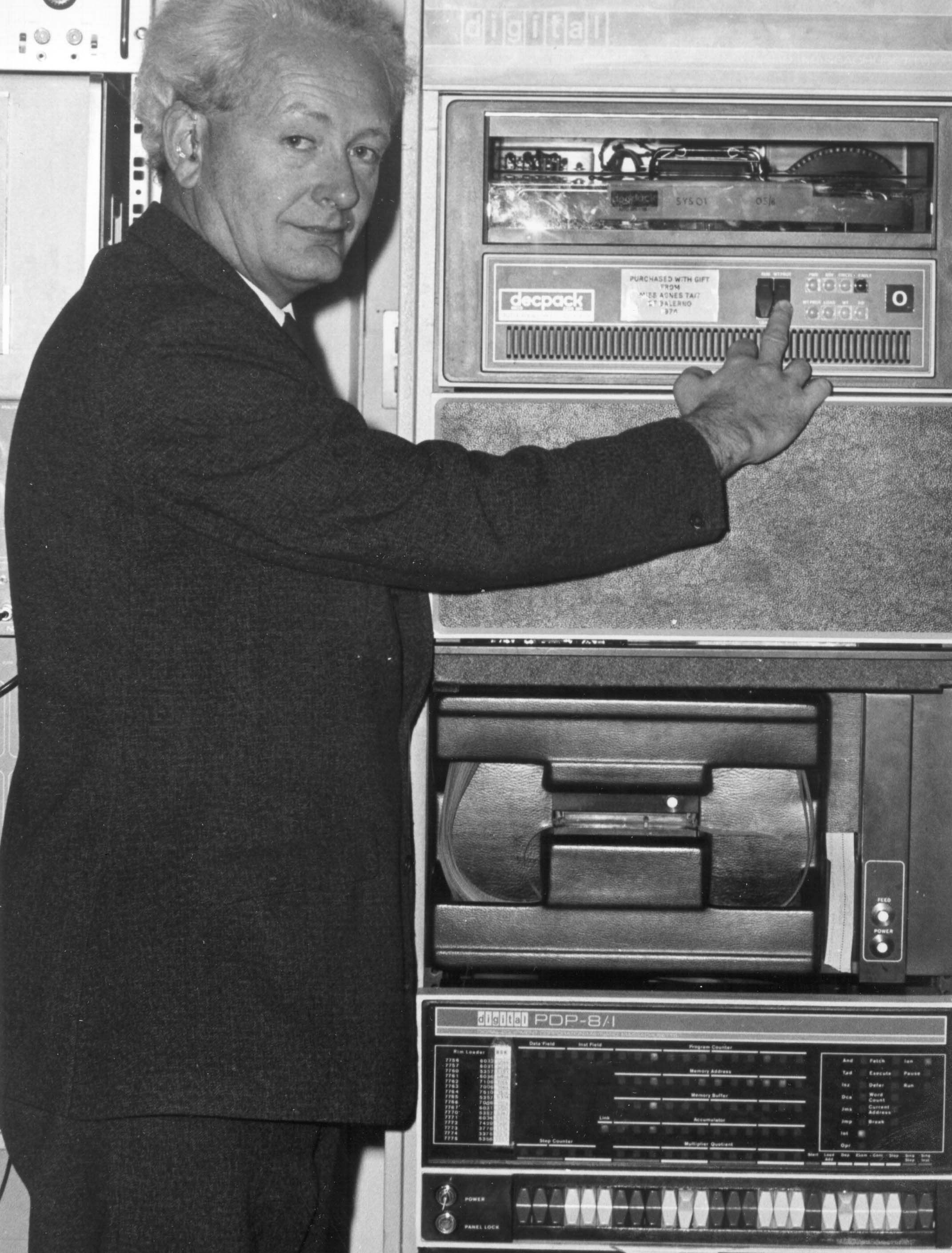

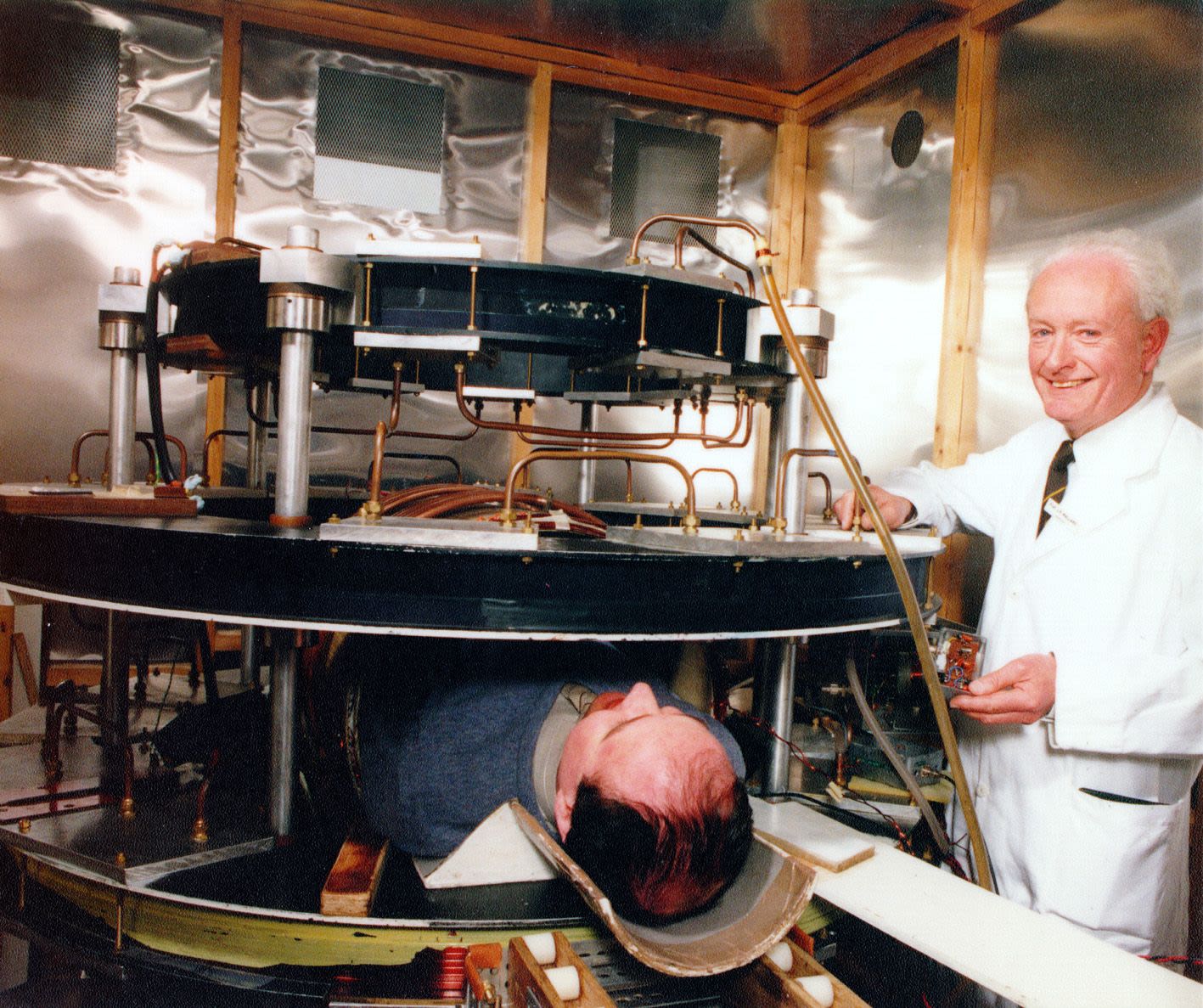
Mallard with the Mark-1 scanner
Mallard with the Mark-1 scanner

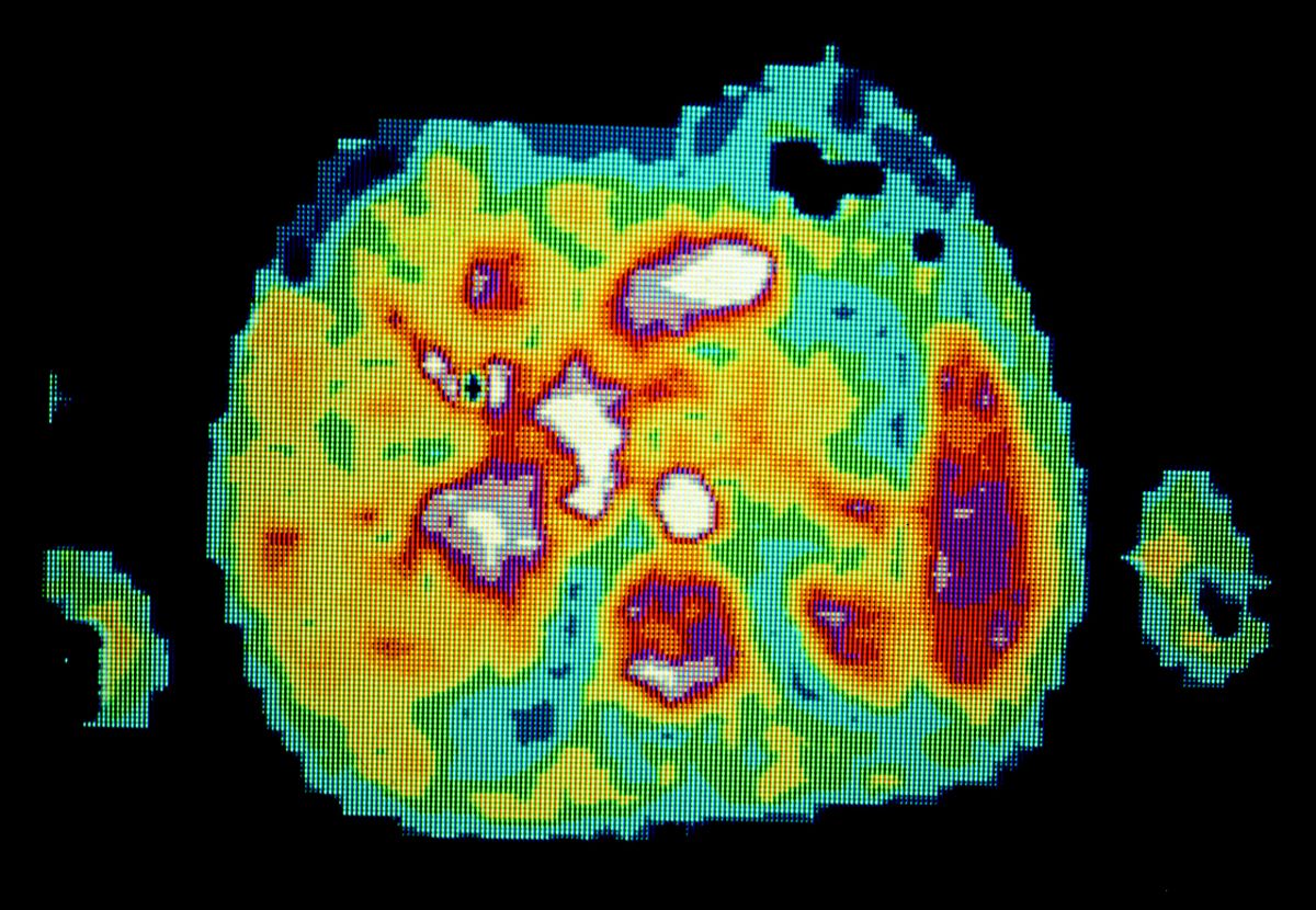
The first MRI patient to be diagnosed in 1980
The first MRI patient to be diagnosed in 1980
Scanning the first patient
Having demonstrated the efficacy of the Mark-1 scanner, they then needed to demonstrate its benefits to real patients.
But reaching this stage required the team to demonstrate its safety and they once again became their own guinea pigs.
Between April and August of 1980, they scanned each other dozens of times to demonstrate that there were no ill effects. Hutchison even diagnosed his own ‘spine warp’ a scoliosis of his upper spine that he had been unaware of and had at first dismissed as a distorted image from the scanner.
By late August, MK-1 was ready for its first real patient. It had been developed on the premise that the relaxation time in malignant disease is different to that of normal tissue and so will shown up in the scans.
But the team needed someone known to have tumours to prove the theory. A man with terminal cancer from Fraserburgh in Aberdeenshire took the bold step of consenting to be the first ever scanned by MRI.
The images showed up clear differences in his liver and spleen and also identified a secondary tumour in his spine which had not previously been known.
It was the breakthrough that Mallard and his team had dedicated more than 20 years to bringing to fruition.
Changing medical treatment throughout the world
As soon as the success of the Mark-1 machine had been demonstrated, a steady stream of patients were brought over from Aberdeen Royal infirmary to the Medical Physics Department where the scanner was located.
It was so popular that the medical soap store in the building had to be hastily reconfigured to create a waiting room for patients.
Just six weeks after its success had been demonstrated in cancer patients, the Mark-1 scanner was also used to capture the first ever images of multiple sclerosis and within two years it had been used in research to identify the effects of alcohol on the brain and the beginnings of dementia.
But such was the popularity of the machine in patient care that the team were no longer able to access the scanner to develop their research.
They sought funding for a Mark-2 machine that would be dedicated to imaging patients but were unsuccessful in the UK and it was only after delivering a lecture in Japan that the country’s technology giants began to take notice.
After securing funding from a Japanese company, a team of four engineers was dispatched to Aberdeen to watch the construction of the second imager and, each day, they faxed their findings back to Japan.
Even after it was taken up in Japan, the team were still met with a great deal of scepticism and the NHS was much slower to adopt its use than other countries such as the USA and Germany.
It was not until the early 1990s that its use started to become more widespread but for younger members of the research team, it limited their UK-based opportunities and many travelled abroad to further their careers.
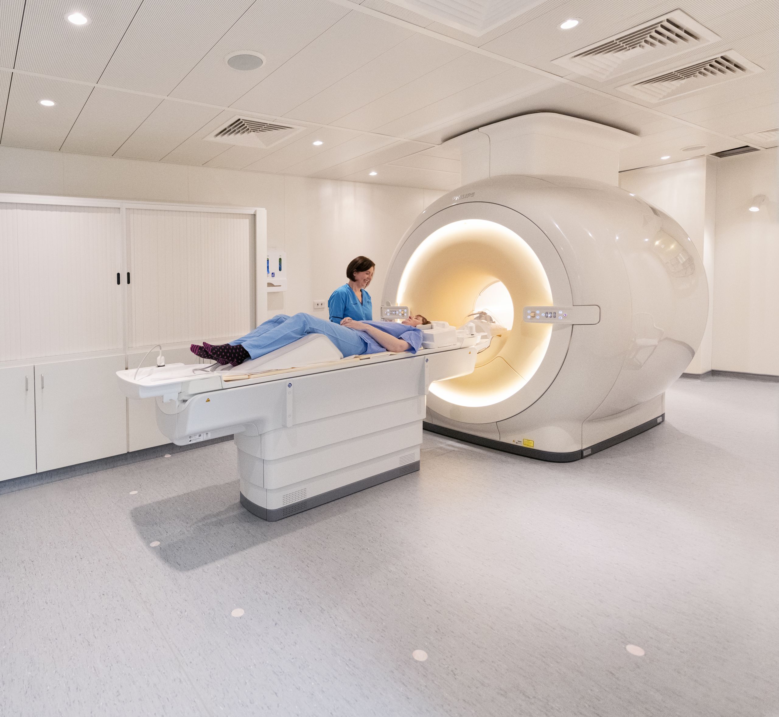
Today MRI machines are a routine part of medical treatment in specialties from psychiatry and cardiology to orthopedics and oncology, allowing clinicians to look inside the human body in amazing clarity and detail.
And although the technology has advanced significantly from the Mark-1 machine built by Mallard’s "small team of half-mad scientists", the spin-warp technique they pioneered still contributes to medical treatment today.
Mallard also continued to champion other advances in imaging technology including the development of PET ( Positron emission tomography), a technique which at the start of his Aberdeen career in 1965 he had predicted "would become one of the most powerful tools for studying human diseases".
His assertion proved correct and it was Mallard who brought Scotland's first PET facility to Aberdeen by leading a national fundraising campaign to pay for a building next to Woodend Hospital to house a second-hand scanner which he had negotiated from researchers in London.
The old facility at Woodend has long since been replaced by the state-of-the-art John Mallard Scottish Pet Centre at Aberdeen Royal Infirmary which at the time was the only PET centre in Scotland.

A lasting Aberdeen legacy
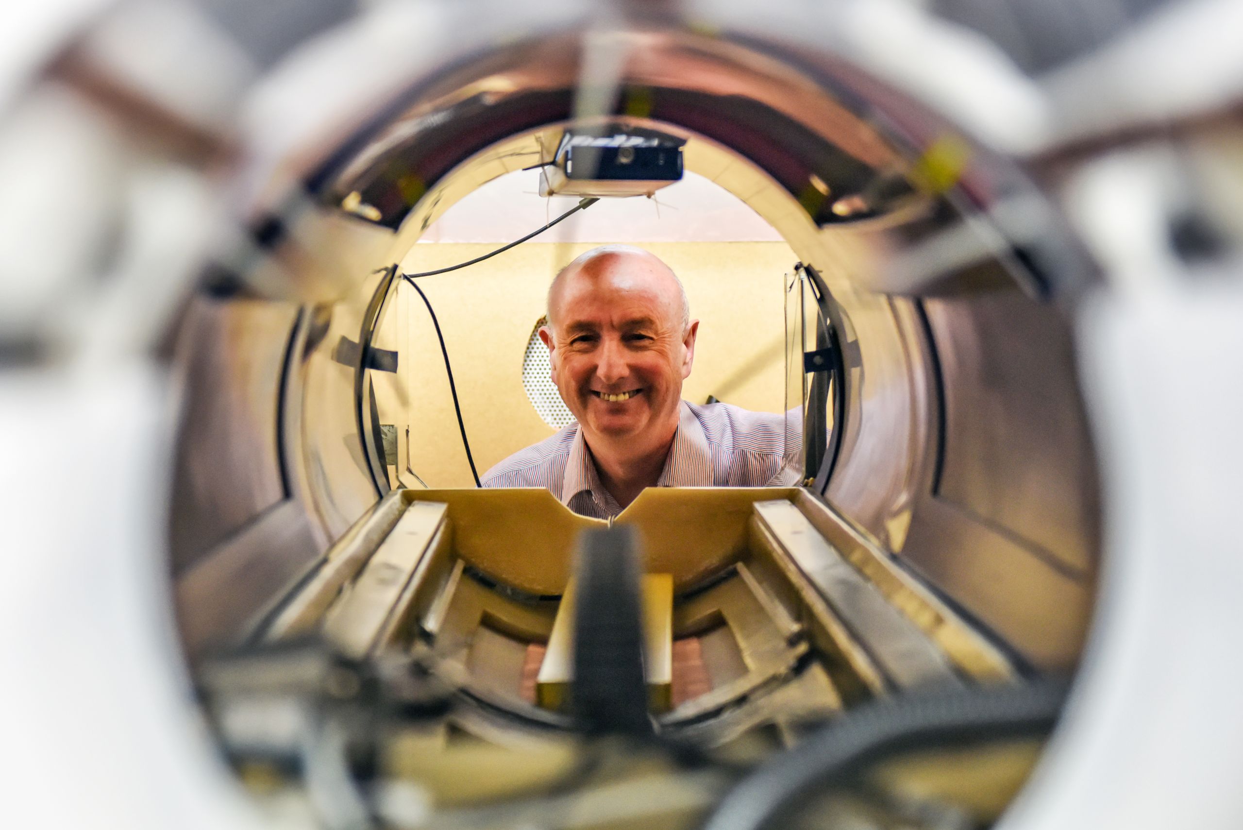
A new-generation “Fast Field-Cycling” MRI scanner is currently being developed at the University’s School of Medicine.
Fast Field Cycling scanners are able to extract much more information by switching the strength of the magnetic field during the scanning procedure and the first ever patients were scanned using FFC in 2017.
Research group leader, Professor David Lurie began his career as a ‘summer student’ in Mallard’s department. He said: “Because FFC scanners can switch their magnetic field, it is almost like having 100 different MRI scanners in one. This gives an extra dimension to the data collected from each patient, greatly expanding the diagnostic potential.
“We are taking major steps towards our technology being adopted by hospitals to benefit patients, which is the ultimate goal of our research.”
The Aberdeen team behind FFC-MRI is leading a 9-strong consortium of research groups from 6 different countries, in a project called “IDentIFY” which received a €6.60 million Horizon 2020 research grant from the European Union to develop the imaging technology and bring it closer to widespread use in hospitals.

Emeritus Professor Peter Sharp, who worked for Professor Mallard and then became his successor, describes his lasting legacy.
“John Mallard was one of those scientists blessed with a vision, a vision of what physics could contribute to healthcare. Many millions of patients worldwide have good reason to be grateful to him.”