Anatomy Collection
With the oldest items in the collection dating to at least the early 19th century, the collection is the product of the research and teaching activities of staff in Aberdeen and includes some 500 specimens and 400 anatomical models, including a life-sized papier-mâché model of a man dating from 1879 by Auzoux.
The collection of 900 works on paper include late 19th century watercolours of anatomical dissections, some of which can still be matched with their original associated wet specimens prepared, and a collection of anatomical drawings produced for Professor Robert Lockhart's Anatomy of the Human Body by seven artists and photographers including Alberto Morrocco.
Access to, and display of, much of the collection is restricted by the Anatomy Act (1984) as amended by the Human Tissue (Scotland) Act 2006.
- About the Anatomy Collection
-
.jpg)
The Anatomy Museum provides a key source for the study of medicine and along with the Pathology and Forensic Medicine Collection, forms the core of the University's medicine and health resources. The University's medical collections are of national importance, providing a comprehensive reference for normal anatomical conditions, which covers the range of functional body systems. The collection totals around 2000 specimens and objects. This includes fluid-preserved specimens of human tissue, osteological material, historical anatomical models, works on paper and a selection of other material used in the preparation of specimens and relating to 19th century grave robbing.
Many of the specimens are preparations of notable past members of staff, such as Professors Struthers (1823-1899), Reid (1851-1939) and Lockhart (1894-1987). Likewise, many of the objects directly relate to the teaching and research interests of such persons. Supporting contextual information is held in the form of past departmental and personal papers and committee minutes at the University's Special Libraries and Archives and provides a rich historical angle to the physical collections.
The historical model collection dates from the late 19th and early 20th centuries. It includes examples from the major model makers of the time, including Auzoux, Maison Tramond, Studio Ziegler and Bock Lips Steger. The models are representative of the media being used at the time with examples in wax, papier-mâché and plaster. Of particular note within the collection for its rarity is a life-sized papier-mâché model of a man dating from 1879 by Auzoux which breaks down into 92 individual pieces. It is one of only a few surviving examples in the world. The anatomical models form an important aspect of the University's wider model collection (held also by the Pathology and Forensic Medicine Collection and the Zoology Museum).
Works on paper form a key feature of the collection. Of particular importance are twenty watercolours of anatomical dissections dating from the early 1890s. What makes them unique is that sixteen can still be matched to their original associated fluid-preserved specimens prepared by Professor Robert Reid in 1890. Whilst it was not uncommon for 19th century anatomy schools to employ an artist, no other such watercolours exist in other Scottish collections whereby they can still be set alongside and compared with their original specimen. Another significant aspect of the works on paper is a collection of anatomical drawings produced for Professor Robert Lockhart's influential 1959 textbook Anatomy of the Human Body (Lockhart et al, 1959). Seven artists and photographers contributed illustrations - Alberto Morrocco, DJ Stephen, W Cruickshank, DW Cameron, RW Matthews, Eric Naylor and Alexander Cain. Alberto Morrocco (1917-1998) is the most prominent of these. Whilst he is best-known for his landscapes of Scotland and Venice, his anatomical work for Lockhart was produced in the post-war period following his service as a conscientious objector in the Medical Corp during WWII. He is not known to have produced any other such anatomical works during his life and this makes our collection an incredibly rare and unique example of his work.
- Past Professors of Anatomy at the University of Aberdeen
-
Chairs of Anatomy
Allen Thomson -1839-1841
Allen Thomson was appointed to the first Chair of Anatomy at Marischal College in 1839 following several years as a lecturer in Anatomy and as a physician in Edinburgh. After his resignation in 1841 he resumed his Anatomy teaching in Edinburgh. In 1842 he became Professor of the Institutes of Medicine at Edinburgh. In 1848 he was appointed as Chair of Anatomy at Glasgow University where he spent the next 29 years. His career was especially distinguished in histology and embryology.
Alexander Jardine Lizars -1841-1863
Allan Jardine Lizars became the first Chair of Anatomy at the University of Aberdeen, formed in 1860 with the joining of Marischal and Kings Colleges. He was forced to retire from his post at the University due to his dipsomania.
1860 Marischal and Kings Colleges join to form the University of Aberdeen
Regius Chairs of Anatomy
Sir John Struthers 1863-1889
Sir John Struthers was a surgeon at Edinburgh Royal Infirmary before joining the University of Aberdeen in 1863 as Chair of Anatomy, a position he was to hold for the next 23 years. He built up a museum of anatomy and introduced the Struthers Medal and Prize. He taught anatomy using the comparative perspective and many of his preparations still exist, both within the Anatomy and Zoology Museums. He retired to Edinburgh in 1889.
Robert Reid 1889-1925
Robert Reid succeeded Struthers to the Regius Chair of Anatomy in 1889. During his career achieved prominence through the discovery of 'Reid's Base Line', and during his time in Aberdeen he made significant contributions to the development of Anatomy and Anthropology in the University and locally. He established an anthropometrical laboratory in the Anatomy Department in 1896, which formed the foundations of the Department's significant work on the growth of children. In 1907, he succeeded in bringing together the disparate collections of anatomical, archaeological and anthropological material that then existed within the University to form the University of Aberdeen Anthropological Museum for which he remained its Honorary Curator until 1937.
Alexander Low 1925-1938
Alexander Low began working for the University in 1894 as assistant and lecturer in the Department of Anatomy following his graduation. He rose to the position of Regius Professor of Anatomy in 1925. His early work on the development of the lower jaw earned him an international reputation, which was strengthened by on-going anthropological research, in particular his detailed and meticulous work on human growth. He laid the foundations for the highly regarded Aberdeen Growth Study of 1956.
Robert Lockhart 1938-1964
Robert Lockhart came to Aberdeen from the University of Birmingham to succeed Alexander Low in 1938 as Regius Professor of Anatomy. With his appointment he also gained the position as Honorary Curator of the Anthropological Museum. He published in the field and his book Anatomy of the Human Body (1959) became a standard text for the study of anatomy.
David Sinclair 1964-1977
David Sinclair joined the University in 1964 as Regius Chair of Anatomy. He made important contributions to curricular development of the anatomical sciences in Aberdeen and he instituted the annual memorial service in 1965 for those who donated their bodies to medical research.
E. John Clegg 1977-1991
E. John Clegg was an acknowledged authority on the historical demography of isolated populations.
Vacant 1991-2019
Simon Parson 2018-present
The current holder of the Regius Chair is Simon Parson. He was appointed Chair in Anatomy at the University of Aberdeen in 2013. His research is focussed on Spinal Muscular Atrophy (SMA), a young-onset form of motor neurone disease.
Pathology and Forensic Medicine Collection
The collection is a unique historical record of disease manifestations in the North-East Scotland in the mid 20th century and includes a few artefacts relating to crimes of note committed in the Aberdeen area around the same period. There are around 1800 fluid-preserved specimens showing both pathological conditions and traumatic pathology, many with associated anonymous clinical case files and supporting contextual material such as weapons and photographic evidence.
The collection also contains late 19th/early 20th century wax, papier-mâché and ceramic models demonstrating a range of pathological appearances, scientific instruments used in the preparation and examination of pathological specimens and associated archival and photographic material. Access to, and display of, parts of the collection is restricted by the Human Tissues Act (1961), as amended by the Human Tissue (Scotland) Act 2006.
- About the Pathology and Forensic Medicine Collection
-

The collection provides an important, comprehensive and unique 20th century record of disease manifestations and traumatic pathology. The collection is one of very few that still remains across the country and it represents the work of a succession of Professors of Pathology and Professors of Forensic Medicine in the University of Aberdeen. It comprises more than 2000 specimens and objects. The collection's strength lies in its specimens of human tissue (around 1800 in total) showing pathological conditions. These are presented in preservative fluid and sealed in Perspex containers. The specimens provide a comprehensive coverage of the range of functional body systems, demonstrating the features of disease in each. The specimens have strongly associated contextual information in the form of anonymous clinical case files that allow the pathology to be viewed in the appropriate clinical settings.
The Forensic Medicine aspect of the collection shows examples of traumatic pathology relating to crimes of note committed in the Aberdeen area. The specimens are presented in the same way as the pathological ones. In many cases, specimens are accompanied by supporting contextual material such as weapons and photographic evidence and complement other forensic collections.
In addition to the fluid-preserved specimens, the collection also includes important wax, papier-mâché and ceramic models dating from the late 19th and early 20th centuries. A number of prominent model makers feature in the collection, including Auzoux of France, Bock Lips Steger of Germany and Towne of London. These models present a variety of pathological appearances and form an important aspect of the University's wider anatomical model collection (held also by the Anatomy and Zoology Museums).
Unique items within the collection that attract interest include a skull in old age used in the 1991 Hollywood production of Hamlet starring Mel Gibson, a small box of thin sections of comparative pathology prepared by Professor of Forensic Medicine Matthew Hay (1855-1932) and a skull with exit bullet wound from the collection of Professor of Surgery Alexander Ogston (1844-1929).
In addition to the specimens and their clinical case files, there is supporting material which provides additional context. This includes several cans of 16mm film made of in vivo experiments and the work-books and notes of past staff. There is also a collection of scientific instruments (mainly microscopes and microtomes) used in the preparation and examination of pathological specimens. In addition, the University's Special Libraries and Archives has a wealth of manuscripts of personal and research papers of past staff and course guides and notes. Associated clinical case files were created between the 1930s-1950s as the Pathology Museum became established. They allow the pathology of the specimens to be viewed in the appropriate clinical settings and a project has been digitising them.
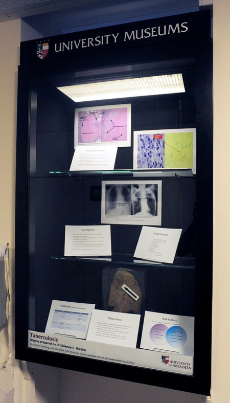
The collection is of significant historic value. It represents an important snapshot of disease and unnatural death during the 20th century, with particular emphasis on the middle third of the century. It provides a unique opportunity to study disease states and traumatic pathology that afflicted the population of the northern Scotland at the time. The fact that some disease conditions represented are no longer prevalent adds to its importance, for example tuberculosis, syphilis and other infectious diseases as the opportunity to collect similar material no longer exists.
