We have already seen in the previous two sections that the Rhynie chert does not crop out anywhere naturally at the surface and thus there are only three main ways of collecting material and/or seeing the chert in situ:
- Collecting loose blocks of chert brought to the surface by ploughing.
- Taking cores of the bed rock by drilling.
- Excavating trenches into the bed rock.
Collecting material and/or seeing the chert in situ
- Trenching at Rhynie
-
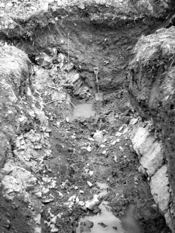
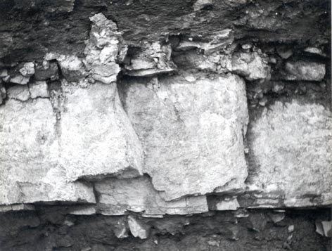
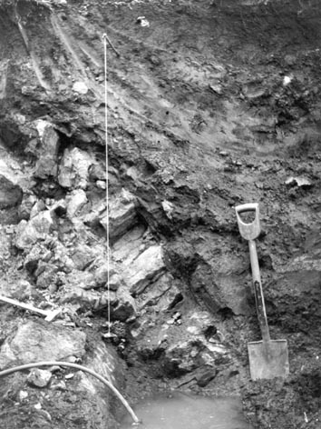
The Rhynie chert is nowhere exposed naturally at the surface, being found primarily as loose 'float' blocks in the fields. Apart from drilling, the only means available to see the chert and interbedded sediments in situ is to dig trenches. The photograph on the left illustrates one of a series of trenches excavated in August 1963. Here the trench has intersected one of the chert beds (the light coloured rock just above left of center and bottom right) where it is interbedded mainly with dark shales and minor muddy sandstones. The beds are dipping quite steeply to the northeast (right as viewed).
By observing the cherts in situ we can better understand their geometry (e.g. whether they occur as tabular beds or lenses), lateral continuity, and distribution in relation to the sedimentary rocks in which they occur.
In 1997, trenching to the north-east of the Rhynie chert locality in the vicinity of Windyfield Farm revealed the only known in situ occurrence of the Windyfield chert (see section on Geology and Setting).
More recent trenching, since 2000, in the northern part of the Rhynie Basin away from the Rhynie chert locality has helped to constrain the sedimentary sequence, stratigraphy and structural geology of the area (see Rice et al. 2002; Rice & Ashcroft in press).
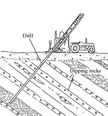
- Drilling at Rhynie
-
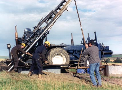
One of the methods employed to sample the Rhynie chert together with the interbedded sedimentary rocks is to 'core' the rocks beneath the surface using a drilling rig (see inset above). If it is known, from previous work, the orientation in which the sedimentary rocks in the subsurface are tilting or 'dipping', the drill can be set at an angle that will be at or near to 900 to the dip of the rocks. This allows the maximum coverage of the sedimentary sequence cored (see inset below).
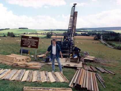
Once recovered, the 'cores' of rock are marked with the respective drilling depth and are then transferred (in depth order) into specially designed boxes for storage and future study (see inset above right).
Other reasons for drilling?
From studying the cores of rock we can analyse not only the chert beds that may be present, but also the sedimentary rocks between the chert beds. In studying various aspects of the sedimentology we can assess many things, from the environment in which the sediments were deposited, to what happened to them after they were buried.
Drilling results help us to understand the stratigraphy of the sediments in the area - both in terms of the age and the 'sequence' in which the sediments were deposited. Another useful aspect of drilling a series of cores over a defined area, is that if we encounter the same chert beds within successive coring operations we can also assess the distribution or lateral continuity of the cherts over that area - in other words where individual chert beds occur and where they die out.
Over the years many cores have been taken through the sedimentary sequence at various localities in the Rhynie area. The drilling projects have provided hundreds of metres of rock core, much of it from the Dryden Flags Formation that contains the Rhynie chert. Drilling has also provided information on the other rock units in the area and on the geological structure of the area (Rice et al. 2002) (see the section on the Site Geology and Setting).
We would like to thank Hays Business Services for housing the cores taken from the Rhynie area at their core store in Aberdeen.
Once the chert is collected, a number of analytical techniques can be used depending upon the information that the researcher is looking for. The following describes four of the main methods that have been employed to study, among other things, the fauna, flora, sedimentology and palaeoenvironments of the cherts.
Four main analytical technique methods
- Cut blocks
-
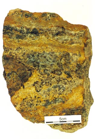
The cut slices of chert can be examined in more detail under reflected light using a binocular microscope after applying a thin veneer of microscopy oil to the cut/polished surface. This enables the identification of, for example, fauna, flora and textures within the chert for more detailed analysis. Further analysis of such features may involve using thin sections, acetate peels and SEM (Scanning Electron Microscope) techniques which are briefly outlined below.
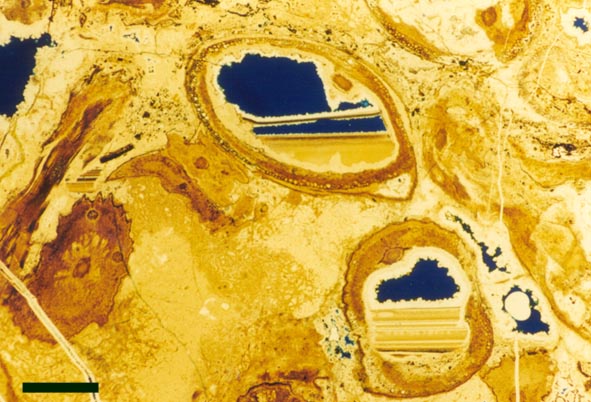
- Thin Sections
-
Thin sections of the rock allow material to be examined under a microscope using transmitted light (i.e.: light that can pass through the rock) as well as reflected light. They are prepared by mounting a small cut and polished sample of chert, using a strong adhesive onto a plate of glass (usually 50x75cm or 25x75cm in size) and then the exposed surface of chert is gradually ground or 'lapped' down to a certain thickness, usually somewhere between 100 and 30μm, depending upon the features being examined. This slice of rock is then usually thin enough to transmit light, enabling a lot more details of fossils and textures to be seen. Thin sections may have a glass cover-slip added for protection of the slide, though alternatively the 'lapped' surface may be polished for examination in back-scattered mode on the SEM (see below).
- Acetate Peels
-
Acetate peels are taken by first etching the cut/polished surface of a chert block with HF (Hydrofluoric acid) for a few seconds. This technique is done under strict safety conditions in a fume cupboard as HF is very dangerous. The acid dissolves away a thin layer of silica leaving a slight positive relief of organic material or any other material that is otherwise insoluble in HF. The etched surface of chert is then flooded with acetone and a sheet of acetate film is then carefully placed over the top and allowed to dry for about 10 minutes. When dry the 'peel' can be pulled from the rock surface and it bears a faithful reproduction of the 'organic' and textural features left in relief after the etching. The peel is then mounted onto a glass slide with a cover-slip for study.
This analytical method has been used extensively in the study of the Rhynie plants and their internal anatomy. Successive peels taken through a plant-bearing block of chert can be helpful in reconstructing the 3D morphology of some of the plants and their growth habit. However, because the features on the peels are often very faint and the peels are not always flat, they are not always so conducive to photography as rock thin sections.
- SEM
-
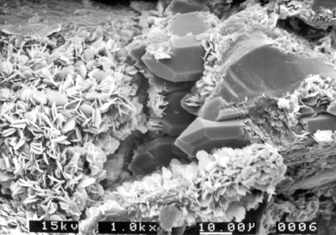
The SEM or Scanning Electron Microscope allows examination of rocks at very high magnifications, showing surface textures and details down to the micron level. This has been a useful technique in looking at many things from clay minerals and diagenetic features in the cherts and associated sediments (see inset above left) to the cuticle surfaces of plants where they have been successfully isolated from the host block of chert.
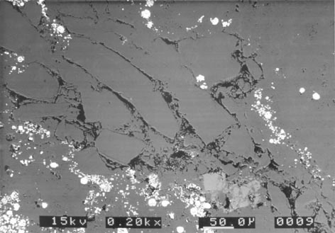
The back-scattered mode on the SEM is particularly useful for looking at textures in polished thin sections of chert and sedimentary rock and highlights differences in mineral chemistries (see inset below left).
- Other Analytical Techniques
-
We have seen four of the techniques employed to study certain aspects of the Rhynie chert, but many other analytical methods are also available to study, for example, the mineralogy and geochemistry of the chert and its associated rocks. Such techniques may include XRD (X-Ray Diffraction), XRF (X-Ray Fluorescence), CL (Cathodo Luminescence) and isotope analysis to name but four. A more detailed review of these and other analytical techniques used to study sedimentary rocks may be found in Tucker (1988).