Microscopy and Histology Core Facility

Microscopy and Histology Core Facility

Explore the hidden world

Microscopy and Histology Core Facility

Services for staff, students and external users

About us

We are pleased to welcome all users with all levels of experience in microscopy and histology to our facility. 

We are located in the Institute of Medical Sciences building at Foresterhill, Aberdeen. 

We have a wide range of modern equipment and techniques available and we are here to help you make the best use of them. We have many years of experience in light, fluorescence and electron microscopy, and histology. We are happy to meet up to help you get started with a new study or help you sort out an unexpected result.

If you are a new user, we can provide support and training on the microscope system you wish to use. 

The Team


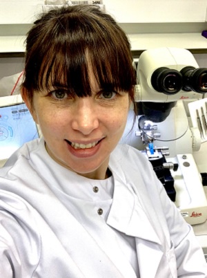
Dr Debbie Wilkinson - Co-Manager and Senior Microscopy Application Specialist 

Photo of Debbie WilkinsonDebbie became a frequent microscope user and an ‘unofficial’ member of the facility in 2004 before officially joining the facility in 2009, after completing her PhD in the Musculoskeletal Research Programme. Over the years Debbie has gained vast amounts of experience in a large variety of microscopy and sample preparation techniques. She can provide training on all the microscopes in the facility and can also deal with sample preparation and training on the transmission and scanning electron microscopes. She is also a proficient user of the two micro CT scanners at the university. She is especially interested in electron tomography and 3D printing. She is keen on public engagement and has taken part in events such as Mayfest. She is also a STEM ambassador and takes part in the STEM Club at Royal Aberdeen Children’s Hospital. She is also an active member and the Aberdeen representative of the Scottish Microscopy Society committee. Debbie is the Laser Protection Supervisor for the IMS and is available to answer any laser safety questions or concerns, she also sits on the IMS Health and Safety committee.

You can find out more information about Debbie on her University of Aberdeen profile.


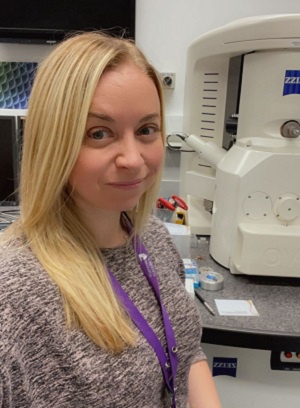
Gillian Milne - Co-Manager and Senior Histology & Electron Microscopy Application Specialist

Photo of Gillian MilneGillian has been part of the technical team at University of Aberdeen since 2003. She started in the Ophthalmology department dealing with samples for histological preparation, then moved over to the newly formed Microscopy and Histology Core Facility in 2006. Here she has gained experience in a variety of histological methods and specialises in transmission electron microscopy. Working with a large variety of sample types (yeast, honey bees, whale, algae to name a few), Gillian has a wealth of knowledge of a number of techniques; high pressure freezing, freeze substitution, microwave processing, resin embedding (for both electron and light microscopy), ultramicrotomy, plus histological techniques; paraffin processing, snap freezing, sectioning and staining. She is also interested in developing techniques for correlative microscopy combining fluorescence and electron microscopy.

You can find out more information about Gillian on her University of Aberdeen profile.


Lucy Wight - Microscopy and Histology Specialist

Photo of Lucy WightLucy joined the facility in 2013, following an MRes at the University of Warwick where she gained experience of confocal and SEM. Prior to this, Lucy completed a BSc Biological Sciences (Microbiology and Infection) at the University of Edinburgh followed by a MSc in Control of Infectious Diseases at the London School of Hygiene and Tropical Medicine. She is experience in providing training on our range of microscopes. She is experienced in sample preparation for fluorescence microscopy and is happy to provide advice regarding this. Lucy can also help with sample preparation for SEM and MicroCT and train users on these systems. Lucy has considerable experience of histology work including cryostat and paraffin processing, embedding, sectioning and histological staining. She is also able to train users in the use of histological equipment such as the wax microtomes, cryostat and staining.

You can find out more information about Lucy on her University of Aberdeen profile.


Dr Natalie Lyall-Varnas - Microscopy Technician

Natalie joined the facility in August 2023 as a microscopy technician and will be here for 1 year to cover maternity leave. Prior to joining the facility Natalie worked in the Netherlands and in England for different biotech companies and carried out her PhD here in Aberdeen Fungal Group. Her project heavily relied on using the confocal spinning disk microscope to image live cells and create videos of transfected macrophages interacting with Candida albicans. She has experience in the preparation of live cells and fixed samples for imaging. She will be training others on various microscopes in the facility including the light, fluorescence, confocal and scanning electron microscopes.


Eilidh Player - Microscopy and Histology Technician

Eilidh joined the Microscopy and Histology facility in April 2023 after working as a technician in a different department at the University of Aberdeen. She first discovered Histology and Microscopy during her master’s project with Dr Vicky Sleight in the Sleight Lab. Eilidh used Histology and Microscopy techniques to investigate Haemocyte cells in slipper snails. In the facility, Eilidh is learning and gaining experience in different histology techniques.

You can find out more about Eilidh on their University of Aberdeen Profile   


Professor Lynda Erskine - Microscopy and Histology Facility Academic Lead and Chair in Development Neurobiology

Lynda is the Academic Lead of the Microscopy and Histology Core Facility. 

You can find out more information about Lynda on her University of Aberdeen profile.


Booking information

All equipment in the facility can be booked using Outlook shared calendars. Each piece of equipment has its own booking calendar (with the exception of microscopes in rooms 3.28/3.29, these rooms have a booking calendar each so you must specify which microscope you want to use within each room when making a booking).


See the following on how to set these up Outlook setup of folders


Note: you may need to wait a few seconds for each calendar to load before you can add a new booking.

An hourly charge is applied for the use of all facility equipment.

Charges

Internal users can access our current charges by clicking on this link.

If you would like a formal quote or if you are an external user, please contact microscopy@abdn.ac.uk.

Acknowledging the Facility

Imaging Facility Guidelines for Acknowledgement

  1. All publications resulting from the use of instruments within the facility should acknowledge the facility as a whole, e.g. ‘the authors gratefully acknowledge the [core facility name] for their support & assistance in this work’ and the facility should be informed of the publication.
  2. Specific grants that have funded the facility instruments used for the work to be published must be acknowledged if the data was acquired during the active period of that grant. Facility staff will advise users of such grant codes.
  3. Assistance above the technical or routine level, with any facility staff providing scientific input and expertise in experimental set-up, acquisition or analysis, should be recognised through co-authorship on resulting publications. Please discuss acknowledgements with facility staff prior to manuscript submission.


Sample preparation

Fast, routine sample preparation with standard protocol.

Simple acknowledgement

Development of new sample preparation protocols. Optimisation of existing protocols for specific samples.

Inclusion of specific facility member on author list

Image acquisition

Training of users to acquire images themselves.

Simple acquisition of data.

Simple acknowledgement

Operational image acquisition with input and decisions dependant on expertise.

Design or re-design of experimental conditions.

Inclusion of specific facility member on author list

Image analysis

Recommendation of analysis software and tools.

Basic data analysis help and advice.

Simple acknowledgement

Constructive data analysis and interpretation.

Creation of complex custom image analysis tools.

Inclusion of specific facility member on author list


Based on the publication policy compiled by Natasha Stephen, Plymouth Electron Microscopy Centre, after discussions with the RMS EM-UK community

Where to find us

We are located on the 1st floor, room 1:58 in the Institute of Medical Sciences building on Foresterhill Campus.

Microscopy and Histology Twitter feed