All are welcome to the official opening of the new Aberdeen Centre for Electron Microscopy, Analysis and Characterisation (ACEMAC) on Thursday 15 February.
There will also be a live demonstration of the new Zeiss GeminiSEM 300 FEG electron microscope.
Images will be beamed from the SEM to the Macrobert Lecture Theatre from 2.30pm to 4pm on the day. There will also be a talk from 2pm to 2.30pm on the Versa XRM CT Scanner by CarlZeiss representatives. The Versa XRM CT Scanner is an instrument that we also have on campus.
On the morning of Friday the 16 February there will be drop-in sessions for researchers to discuss their own samples with microscopy technicians and CarlZeiss representatives. Email acemac@abdn.ac.uk for further details on this. Between 11am and 12pm on Friday the 16 there will be another live demonstration of the electron microscope in the Macrobert Lecture Theatre displaying a different suite of samples from those shown on the Thursday.
Optical teaching microscopes will also be on display, with a demonstration of the CarlZeiss Digital Microscopy Classroom (https://www.zeiss.com/microscopy/int/solutions/education/digital-classroom.html ). This follows a demonstration of digital microscopy for teaching earlier this month by Nikon, and will be followed by demonstrations by Olympus and Leica in the coming weeks.
The CarlZeiss teaching microscopes will be located in Meston 122 and in place from Monday the 12 February until Friday the 23 February. CarlZeiss representatives will be on hand on Thursday and Friday of next week.
Staff interested in trying the Zeiss digital microscopes in live classroom situations are encouraged to do so, and both staff and students are encouraged to try them out at their own convenience, and give their feedback and opinions on the approach and equipment. Contact a.brasier@abdn.ac.uk for further details.
Alex Brasier, Lecturer, Geology & Petroleum Geology, said: “A Scanning Electron Microscope is a kind of microscope that uses electrons rather than light. This enables us to look at very small objects (micro-to nanoscale, i.e. from millimetres to just under 1 billionth of a metre), including metals (for engineering), rocks (in the geosciences), archaeological artefacts, chemicals, insects etc.
It’s a University-wide Core Facility, open to all across the University and to external academic / industrial customers and not specific to any particular subject or discipline.
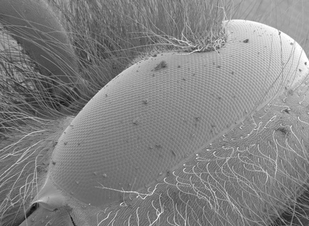
There are a number of different detectors on the microscope. Several are used for high-resolution imaging at the scale of millimetres to nanometres. We can use these, for example, to image the eyes of flies, or grains of sand in a sandstone. Some of the detectors also give compositional information, including an Energy Dispersive Specroscopy (EDS) detector that enables us to produce elemental ‘maps’ of samples. We also have an Electron Backscatter Diffraction (EBSD) detector which helps us identify crystals in our samples, and also gives information on stress and strain that have affected the specimen. This can be used in geology and chemistry for identifying minerals and chemicals, and also in engineering for looking at how different metals perform under stress.”