
Trigonotarbids are an extinct order of terrestrial arachnids related to modern day spiders. The earliest trigonotarbid known in the fossil record is from the Silurian Ludlow Bone Bed (Jeram et al. 1990). It was formally described by Jason Dunlop (1996a). The group ranges from the Late Silurian to the Early Permian; being most diverse in genera and species during the Late Carboniferous. During this time the group occupied a place in some of the earliest terrestrial ecosystems (Rhynie) as well as being an important faunal element of coal swamp communities. Fossil arachnids are relatively rare and only occur in sites of exceptional preservation where their unmineralised cuticle can be fossilised either due to unusual chemical conditions or rapidity of burial. The Lower Devonian trigonotarbids of Rhynie are amongst the most completely preserved and best known members of the group. They are so well preserved that features such as their respiratory organs (book lungs) (Claridge & Lyon 1961), mouthparts (Dunlop 1994) and even muscle tendons have been identified in some sections of the chert.

- Relationships
-
Although sharing a similar body plan with other arachnids, trigonotarbids are not spiders as such. They belong within a group of animals called the chelicerates. Other chelicerates include the horseshoe crabs (such as modern Limulus), scorpions, the extinct eurypterids and all other arachnids including mites. As the earliest recognised terrestrial animal fossil is an Upper Silurian trigonotarbid, understanding of the group obviously plays an important role in determining the timing and mechanism by which animals invaded the land.
There are a number of significant differences between trigonotarbids (informally called "trigs") and spiders which are tabulated below:
Spiders Trigonotarbids Silk producing spinnerets Spinnerets absent Smooth opisthosoma (with the exception of liphistid spiders) Segmented opisthosoma Lateral and median eyes consolidated on a single tubercle Separate lateral and median eye tubercles - Identity
-
Hirst (1923) originally described five species of trigonotarbids from the Rhynie cherts under the generic names Palaeocharinoides and Palaeocharinus. These are Palaeocharinoides hornei, Palaeocharinus rhyniensis, P. scourfieldi, P.calmani and P. kidstoni.
The two genera were distinguished respectively by an acute vs. a straight posterior margin of the sternum (a central plate located on the ventral side of the head region). Shear et al. (1987) rejected this as a diagnostic generic character and synonymised Palaeocharinoides with Palaeocharinus.
The number of species should also be treated with caution as many were diagnosed as different from one another on very minor differences. Some of these apparent differences may have been introduced during the fossilisation process or could be accounted for by intra-specific variation. Of Hirst's trigonotarbid specimens, it would appear only two valid species are probably present: Palaeocharinus rhyniensis and Palaeocharinus hornei.
Recently a new species, Palaeocharinus tuberculatus, has been described from the Windyfield chert (Fayers et al. 2004), differing from Hirst's specimens in its larger size and distinctive tuberculate ornament (see inset below).
The following section gives the general morphology of the trigonotarbids known form the cherts at Rhynie.
- Morphology
-
The Rhynie trigonotarbid arachnids described to date all belong to the family Palaeocharinidae, and possess a distinctive and characteristic morphology by which they can be identified. Overall trigonotarbids consist of a prosoma (head region) and an opisthosoma (body region) (see inset below).
Each of these areas is explored in more detail below. The Rhynie 'trigs' are generally quite small in size. The most commonly encountered species (Palaeocharinus rhyniensis and P. hornei) have a body length of up to 4 mm. Palaeocharinus tuberculatus is slightly larger having a maximum body length of at least 6mm. However, other trigonotarbid specimens have occasionally been found in the cherts with body lengths ranging between 14 mm and 20 mm. These larger forms (currently being described) appear to belong to a new species quite distinct from Palaeocharinus and possibly belong to a separate family.


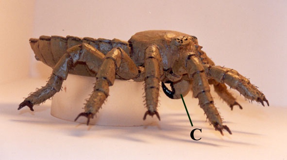
Head Region

Underneath the domed carapace hangs a pair of down and backwardly directed poison fangs or chelicerae; the morphological character from which the group derives its name (see inset below). Towards the posterior of the carapace are the leg attachment points (coxae). The position of the leg appendages, associated with the prosoma is a further diagnostic characteristic of the Chelicerates.
Body Region


Prosomal Appendages
Just as in extant spiders, trigonotarbids possessed a pair of chelicerae (fangs), a pair of pedipalps and four pairs of walking legs. The fangs were used in prey capture and the pedipalps may have had a sensory function, although in extant male spiders they are used in mating. Each of the individual leg segments or podomeres can be named. Starting from the insertion point they are as follows: coxa, trochanter, femur, patella, tibia, basitarsus and telotarsus (see reconstruction above and inset below). In trigonotarbids the most distal of the leg podomeres, the telotarsus, bears a pair of tarsal claws that were an adaptation for walking in the terrestrial environment. The more distal podomeres of the walking legs bear and increasing number of fine setae or hairs. These were used by the animal to augment its sensory array.
The legs of Palaeocharinus tuberculatus differ from the other Palaeocharinus species in that they possess distinctive longitudinal rows of tubercles. Many of these tubercles appear to flank and guard socketed sensory hairs.

- Reconstruction
-


- Palaeoecology
-
Trigonotarbid arachnids such as the Rhynie form Palaeocharinus did not possess the morphological structures termed spinnerets. Extant spiders have spinnerets and these are used to produce silk, one of the most important substances in terms of spider ecology.
Silk threads spun from the spinnerets are used to build food-capturing webs, silk lines the burrows of trap door spiders, is used to wrap prey items and also to provide protective cocoons for the eggs.
Some species even use the silk as a mode of transport. By letting out a long thread of silk into the prevailing wind small spiders 'balloon' and can travel great distances. This can aid widespread dispersal of the species. As trigonotarbids lack this innovation, their possible palaeoecological interpretation must be somewhat limited.
Another group of extant arachnids which provide a more useful analogue to trigonotarbids are the opilionids, or 'harvestmen'. These ground living forms lack silk but instead immobilise their prey with a bite from poisonous fangs (chelicerae). It is conceivable the Rhynie trigonotarbids behaved in a similar manner since they possessed particularly robust and well-developed fangs.

The robust walking legs and downwardly directed fangs suggest that Palaeocharinus may have been a ground predator stalking the micro-habitat provided by the stands of Rhynie plants. It may well have sat in wait and ambushed its prey from the cover of fallen plant stems. Indeed some trigonotarbid fossils have been discovered within the cherts within the hollow stems and sporangia of the Rhynie plants (Kevan et al. 1975) (see inset above). They may have used these as cover from which to ambush their prey. Alternatively such locations may have provided ideal conditions for ecdysis or the moulting process to take place in.